|
Comment citer 1. Pour la citation dans le texte (Matériel & Méthodes) : 2. Pour le tableau des ressources clés : |
||
|
Numéro vert : (877) 796-6397 -- États-Unis et Canada uniquement -- |
Fax : +1-832-582-8590 Commandes : +1-832-582-8158 |
Support technique : +1-832-582-8158 Ext:3 Veuillez indiquer votre numéro de commande dans le-mail. Nous nous efforçons de répondre à toutes les demandes par e-mail dans un délai dun jour ouvrable. |
Description biologique
| Spécificité | Artemis Antibody [H20J21] détecte les niveaux endogènes de la protéine Artemis totale. |
|---|---|
| Contexte | Artemis (DCLRE1C, également connu sous le nom de SNM1C) est une nucléase nucléaire exprimée de manière ubiquitaire appartenant à la super-famille des métallo-β-lactamases (MBL), essentielle pour la réparation de l'ADN par jonction d'extrémités non homologues (NHEJ) et la recombinaison V(D)J dans les lymphocytes en développement. Artemis est une protéine de 692 acides aminés avec un domaine catalytique N-terminal (résidus 1-360) comprenant un repliement MBL central (sandwich α/β-β/α) et un sous-domaine β-CASP intégré contenant les motifs conservés 1-4 (His33, His35, His115, Asp116 coordonnant les ions Zn²⁺ M1/M2) et les motifs A-C pour le traitement des acides nucléiques, ainsi qu'une queue régulatrice C-terminale largement non structurée (résidus 361-692) qui auto-inhibe l'activité et assure l'ancrage de DNA-PKcs. DNA-PKcs phosphoryle Artemis (par exemple, au niveau de Thr127, Ser251) lors de la reconnaissance des DSB, activant son activité endonucléasique structure-spécifique pour ouvrir les extrémités codantes scellées en épingle à cheveux dans la recombinaison V(D)J, couper les surplombs 5'/3', les volets et les bulles dans le NHEJ pour la ligature XRCC4/LIG4, et traiter les extrémités complexes induites par les IR tout en antagonisant la HR via l'interaction 53BP1-PTIP. ATM/ATR phosphorylent davantage Artemis pour la récupération des points de contrôle G2/M et de phase S, favorisant l'activation de Cdk1-cyclin B et la dégradation de la cycline E via SCF^{Fbw7}. Les mutations bialléliques entraînent une immunodéficience combinée sévère radiosensible (RS-SCID) avec un défaut de jonction V(D)J et une instabilité génomique prédisposant à la malignité. |
Informations dutilisation
| Application | WB, IP | Dilution |
|
||||
|---|---|---|---|---|---|---|---|
| Réactivité | Human, Monkey | ||||||
| Source | Rabbit Monoclonal Antibody | MW | 90 kDa | ||||
| Tampon de stockage | PBS, pH 7.2+50% Glycerol+0.05% BSA+0.01% NaN3 | Stockage (À partir de la date de réception) |
-20°C (avoid freeze-thaw cycles), 2 years | ||||
| WB |
Experimental Protocol:
Sample preparation
1. Tissue: Lyse the tissue sample by adding an appropriate volume of ice-cold RIPA/Nuclear Lysis Buffer (containing Protease Inhibitor Cocktail),and homogenize the tissue at a low temperature. 2. Adherent cell: Aspirate the culture medium and wash the cells with ice-cold PBS twice. Lyse the cells by adding an appropriate volume of RIPA/Nuclear Lysis Buffer (containing Protease Inhibitor Cocktail) and put the sample on ice for 5 min. 3. Suspension cell: Transfer the culture medium to a pre-cooled centrifuge tube. Centrifuge and aspirate the supernatant. Wash the cells with ice-cold PBS twice. Lyse the cells by adding an appropriate volume of RIPA/Nuclear Lysis Buffer (containing Protease Inhibitor Cocktail) and put the sample on ice for 5 min. 6. Add protein loading buffer, heat 20 μL of the sample at 95~100°C for 5 min, let it cool down on ice and then centrifuge for 5 min. Electrophoretic separation
1. According to the concentration of extracted protein, load appropriate amount of protein sample and marker onto SDS-PAGE gels for electrophoresis. Recommended separating gel (lower gel) concentration: 10%. Reference Table for Selecting SDS-PAGE Separation Gel Concentrations 2. Power up 80V for 30 minutes. Then the power supply is adjusted (110 V~150 V), the Marker is observed, and the electrophoresis can be stopped when the indicator band of the predyed protein Marker where the protein is located is properly separated. (Note that the current should not be too large when electrophoresis, too large current (more than 150 mA) will cause the temperature to rise, affecting the result of running glue. If high currents cannot be avoided, an ice bath can be used to cool the bath.)
Transfer membrane
1. Take out the converter, soak the clip and consumables in the pre-cooled converter;
2. Activate PVDF membrane with methanol for 1 min and rinse with transfer buffer;
3. Install it in the order of "black edge of clip - sponge - filter paper - filter paper - glue -PVDF membrane - filter paper - filter paper - sponge - white edge of clip"; 4. The protein was electrotransferred to PVDF membrane. ( 0.45 µm PVDF membrane is recommended ) Reference Table for Selecting PVDF Membrane Pore Size Specifications Recommended conditions for wet transfer: 200 mA, 120 min. ( Note that the transfer conditions can be adjusted according to the protein size. For high-molecular-weight proteins, a higher current and longer transfer time are recommended. However, ensure that the transfer tank remains at a low temperature to prevent gel melting.)
Block
1. After electrotransfer, wash the film with TBST at room temperature for 5 minutes;
2. Incubate the film in the blocking solution for 1 hour at room temperature;
3. Wash the film with TBST for 3 times, 5 minutes each time.
Antibody incubation
1. Use 5% skim milk powder to prepare the primary antibody working liquid (recommended dilution ratio for primary antibody 1:1000), gently shake and incubate with the film at 4°C overnight; 2. Wash the film with TBST 3 times, 5 minutes each time;
3. Add the secondary antibody to the blocking solution and incubate with the film gently at room temperature for 1 hour;
4. After incubation, wash the film with TBST 3 times for 5 minutes each time.
Antibody staining
1. Add the prepared ECL luminescent substrate (or select other color developing substrate according to the second antibody) and mix evenly;
2. Incubate with the film for 1 minute, remove excess substrate (keep the film moist), wrap with plastic film, and expose in the imaging system. (Exposure time of at least 60s is recommended)
|
Références
|
Données dapplication
WB
Validé par Selleck
-
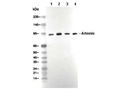 Lane 1: Jurkat, Lane 2: HT-29, Lane 3: PC-3, Lane 4: MCF-7