|
Comment citer 1. Pour la citation dans le texte (Matériel & Méthodes) : 2. Pour le tableau des ressources clés : |
||
|
Numéro vert : (877) 796-6397 -- États-Unis et Canada uniquement -- |
Fax : +1-832-582-8590 Commandes : +1-832-582-8158 |
Support technique : +1-832-582-8158 Ext:3 Veuillez indiquer votre numéro de commande dans le-mail. Nous nous efforçons de répondre à toutes les demandes par e-mail dans un délai dun jour ouvrable. |
Description biologique
| Spécificité | β Amyloid (Aβ) 1-42 Antibody [H15J22] détecte les niveaux endogènes de protéine β Amyloid 1-42 totale. |
|---|---|
| Contexte | Amyloid β 1-42 (Aβ42) est un peptide de 42 acides aminés généré à partir de la protéine précurseur de l'amyloïde (APP) par clivage séquentiel par la β-sécrétase et la γ-sécrétase dans les neurones. Structurellement, Aβ42 est fortement hydrophobe à son C-terminus, ce qui favorise un changement conformationnel d'α-hélice en feuillet β qui facilite l'agrégation en oligomères solubles, protofibrilles et fibrilles amyloïdes insolubles. Aβ42 est exprimé principalement dans le cerveau, en particulier dans le néocortex, et est plus sujet à l'agrégation et plus neurotoxique que la forme plus courte Aβ40. Fonctionnellement, l'accumulation pathologique d'Aβ42 – en particulier sous forme d'oligomères solubles – est fortement impliquée dans la pathogenèse de la maladie d'Alzheimer en perturbant la signalisation synaptique, en altérant la mémoire et en contribuant à la formation de plaques. |
Informations dutilisation
| Application | IHC | Dilution |
|
||
|---|---|---|---|---|---|
| Réactivité | Mouse, Rat, Human | ||||
| Source | Rabbit Monoclonal Antibody | MW | |||
| Tampon de stockage | PBS, pH 7.2+50% Glycerol+0.05% BSA+0.01% NaN3 | Stockage (À partir de la date de réception) |
-20°C (avoid freeze-thaw cycles), 2 years | ||
| IHC |
Experimental Protocol:
Deparaffinization/Rehydration
1. Deparaffinize/hydrate sections:
2. Incubate sections in three washes of xylene for 5 min each.
3. Incubate sections in two washes of 100% ethanol for 10 min each.
4. Incubate sections in two washes of 95% ethanol for 10 min each.
5. Wash sections two times in dH2O for 5 min each.
6.Antigen retrieval: For Citrate: Heat slides in a microwave submersed in 1X citrate unmasking solution until boiling is initiated; continue with 10 min at a sub-boiling temperature (95°-98°C). Cool slides on bench top for 30 min.
Staining
1. Wash sections in dH2O three times for 5 min each.
2. Incubate sections in 3% hydrogen peroxide for 10 min.
3. Wash sections in dH2O two times for 5 min each.
4. Wash sections in wash buffer for 5 min.
5. Block each section with 100–400 µl of blocking solution for 1 hr at room temperature.
6. Remove blocking solution and add 100–400 µl primary antibody diluent in to each section. Incubate overnight at 4°C.
7. Remove antibody solution and wash sections with wash buffer three times for 5 min each.
8. Cover section with 1–3 drops HRPas needed. Incubate in a humidified chamber for 30 min at room temperature.
9. Wash sections three times with wash buffer for 5 min each.
10. Add DAB Chromogen Concentrate to DAB Diluent and mix well before use.
11. Apply 100–400 µl DAB to each section and monitor closely. 1–10 min generally provides an acceptable staining intensity.
12. Immerse slides in dH2O.
13. If desired, counterstain sections with hematoxylin.
14. Wash sections in dH2O two times for 5 min each.
15. Dehydrate sections: Incubate sections in 95% ethanol two times for 10 sec each; Repeat in 100% ethanol, incubating sections two times for 10 sec each; Repeat in xylene, incubating sections two times for 10 sec each.
16. Mount sections with coverslips and mounting medium.
|
Références
|
Données dapplication
IHC
Validé par Selleck
-
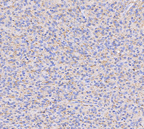 Immunohistochemical analysis of formalin fixed paraffin embedded human brain tissue with F3375 at 1:1000 dilution.