|
Comment citer 1. Pour la citation dans le texte (Matériel & Méthodes) : 2. Pour le tableau des ressources clés : |
||
|
Numéro vert : (877) 796-6397 -- États-Unis et Canada uniquement -- |
Fax : +1-832-582-8590 Commandes : +1-832-582-8158 |
Support technique : +1-832-582-8158 Ext:3 Veuillez indiquer votre numéro de commande dans le-mail. Nous nous efforçons de répondre à toutes les demandes par e-mail dans un délai dun jour ouvrable. |
Description biologique
| Spécificité | Cas9 (S. pyogenes) Antibody [L14C20] détecte les niveaux endogènes de la protéine Cas9 (S. pyogenes) totale. |
|---|---|
| Contexte | La Cas9 de Streptococcus pyogenes (SpCas9) est une endonucléase guidée par ARN de 1 368 acides aminés, centrale à l'immunité adaptative CRISPR de type II, formant un complexe ribonucléoprotéique avec un ARN guide (gRNA : hybride crRNA-tracrRNA) qui reconnaît et clive l'ADN viral envahisseur au niveau des sites NGG protospacer adjacent motif (PAM), une propriété qui a fait de SpCas9 un outil fondamental pour les applications d'édition génomique précise. SpCas9 adopte une architecture bilobée : un lobe de reconnaissance N-terminal (REC) composé des domaines REC1 et REC2 qui berce le gRNA pour le balayage de la cible, et un lobe de nucléase C-terminal (NUC) abritant les domaines endonucléases RuvC (résidus 1–59, 718–769, 1096–1184) et HNH (résidus 775–908), séparés par une hélice de pont hélicoïdale interrogeant le PAM (Arg1333/Glu1219). Des lobes structuraux L1 et L2 supplémentaires stabilisent le canal central à travers lequel l'ADN s'enfile après l'appariement d'amorces. Le complexe gRNA-Cas9 scanne initialement l'ADN pour les séquences PAM via de légers contacts dans le petit sillon (Arg1335/Gln1119), puis initie l'appariement des bases à partir de la région « amorce » proximale au PAM (positions 1–12), déplaçant le brin d'ADN non-cible pour former un hybride en boucle R stabilisé par un réseau de liaisons hydrogène (notamment Ser808/Arg661 avec le squelette phosphate). Cela déclenche un changement conformationnel qui ouvre les centres catalytiques HNH et RuvC, chacun coordonné par deux ions Mg²⁺ : HNH (Arg661/Asp839) clive le brin cible, et RuvC (Asp10/His983/Asp986) coupe le brin non-cible, ce qui entraîne une cassure double brin (DSB) franche à 3 pb en amont du PAM via un clivage séquentiel (RuvC avant HNH, avec un délai d'environ 0,2 s). La fidélité est assurée par la déstabilisation des mésappariements d'amorces et la relecture distale au PAM. La nature programmable de SpCas9 permet non seulement l'édition génomique, mais aussi l'édition de bases (par exemple, les fusions dCas9-cytosine désaminase) et la régulation transcriptionnelle (par exemple, dCas9-VP64 pour l'activation/répression), avec des applications thérapeutiques telles que la correction de la drépanocytose par l'édition de BCL11A. |
Informations dutilisation
| Application | WB, IF, FCM | Dilution |
|
||||||
|---|---|---|---|---|---|---|---|---|---|
| Réactivité | All Species Expected | ||||||||
| Source | Mouse Monoclonal Antibody | MW | 160 kDa | ||||||
| Tampon de stockage | PBS, pH 7.2+50% Glycerol+0.05% BSA+0.01% NaN3 | Stockage (À partir de la date de réception) |
-20°C (avoid freeze-thaw cycles), 2 years | ||||||
| WB |
Experimental Protocol:
Sample preparation
1. Tissue: Lyse the tissue sample by adding an appropriate volume of ice-cold RIPA/NP-40 Lysis Buffer (containing Protease Inhibitor Cocktail),and homogenize the tissue at a low temperature. 2. Adherent cell: Aspirate the culture medium and wash the cells with ice-cold PBS twice. Lyse the cells by adding an appropriate volume of RIPA/NP-40 Lysis Buffer (containing Protease Inhibitor Cocktail) and put the sample on ice for 5 min. 3. Suspension cell: Transfer the culture medium to a pre-cooled centrifuge tube. Centrifuge and aspirate the supernatant. Wash the cells with ice-cold PBS twice. Lyse the cells by adding an appropriate volume of RIPA/NP-40 Lysis Buffer (containing Protease Inhibitor Cocktail) and put the sample on ice for 5 min. 4. Place the lysate into a pre-cooled microcentrifuge tube. Centrifuge at 4°C for 15 min. Collect the supernatant;
5. Remove a small volume of lysate to determine the protein concentration;
6. Combine the lysate with protein loading buffer. Boil 20 µL sample under 95-100°C for 5 min. Centrifuge for 5 min after cool down on ice.
Electrophoretic separation
1. According to the concentration of extracted protein, load appropriate amount of protein sample and marker onto SDS-PAGE gels for electrophoresis. Recommended separating gel (lower gel) concentration: 5%. Reference Table for Selecting SDS-PAGE Separation Gel Concentrations 2. Power up 80V for 30 minutes. Then the power supply is adjusted (110 V~150 V), the Marker is observed, and the electrophoresis can be stopped when the indicator band of the predyed protein Marker where the protein is located is properly separated. (Note that the current should not be too large when electrophoresis, too large current (more than 150 mA) will cause the temperature to rise, affecting the result of running glue. If high currents cannot be avoided, an ice bath can be used to cool the bath.)
Transfer membrane
1. Take out the converter, soak the clip and consumables in the pre-cooled converter;
2. Activate PVDF membrane with methanol for 1 min and rinse with transfer buffer;
3. Install it in the order of "black edge of clip - sponge - filter paper - filter paper - glue -PVDF membrane - filter paper - filter paper - sponge - white edge of clip"; 4. The protein was electrotransferred to PVDF membrane. ( 0.45 µm PVDF membrane is recommended ) Reference Table for Selecting PVDF Membrane Pore Size Specifications Recommended conditions for wet transfer: 200 mA, 120 min. ( Note that the transfer conditions can be adjusted according to the protein size. For high-molecular-weight proteins, a higher current and longer transfer time are recommended. However, ensure that the transfer tank remains at a low temperature to prevent gel melting.)
Block
1. After electrotransfer, wash the film with TBST at room temperature for 5 minutes;
2. Incubate the film in the blocking solution for 1 hour at room temperature;
3. Wash the film with TBST for 3 times, 5 minutes each time.
Antibody incubation
1. Use 5% skim milk powder to prepare the primary antibody working liquid (recommended dilution ratio for primary antibody 1:1000), gently shake and incubate with the film at 4°C overnight; 2. Wash the film with TBST 3 times, 5 minutes each time;
3. Add the secondary antibody to the blocking solution and incubate with the film gently at room temperature for 1 hour;
4. After incubation, wash the film with TBST 3 times for 5 minutes each time.
Antibody staining
1. Add the prepared ECL luminescent substrate (or select other color developing substrate according to the second antibody) and mix evenly;
2. Incubate with the film for 1 minute, remove excess substrate (keep the film moist), wrap with plastic film, and expose in the imaging system.
|
| IF |
Experimental Protocol:
Sample Preparation
1. Adherent Cells: Place a clean, sterile coverslip in a culture dish. Once the cells grow to near confluence as a monolayer, remove the coverslip for further use.
2. Suspension Cells: Seed the cells onto a clean, sterile slide coated with poly-L-lysine.
3. Frozen Sections: Allow the slide to thaw at room temperature. Wash it with pure water or PBS for 2 times, 3 minutes each time.
4. Paraffin Sections: Deparaffinization and rehydration. Wash the slide with pure water or PBS for 3 times, 3 minutes each time. Then perform antigen retrieval.
Fixation
1. Fix the cell coverslips/spots or tissue sections at room temperature using a fixative such as 4% paraformaldehyde (4% PFA) for 10-15 minutes.
2. Wash the sample with PBS for 3 times, 3 minutes each time.
Permeabilization
1.Add a detergent such as 0.1–0.3% Triton X-100 to the sample and incubate at room temperature for 10–20 minutes.
(Note: This step is only required for intracellular antigens. For antigens expressed on the cell membrane, this step is unnecessary.)
Wash the sample with PBS for 3 times, 3 minutes each time.
Blocking
Add blocking solution and incubate at room temperature for at least 1 hour. (Common blocking solutions include: serum from the same source as the secondary antibody, BSA, or goat serum.)
Note: Ensure the sample remains moist during and after the blocking step to prevent drying, which can lead to high background.
Immunofluorescence Staining (Day 1)
1. Remove the blocking solution and add the diluted primary antibody.
2. Incubate the sample in a humidified chamber at 4°C overnight.
Immunofluorescence Staining (Day 2)
1. Remove the primary antibody and wash with PBST for 3 times, 5 minutes each time.
2. Add the diluted fluorescent secondary antibody and incubate in the dark at 4°C for 1–2 hours.
3. Remove the secondary antibody and wash with PBST for 3 times, 5 minutes each time.
4. Add diluted DAPI and incubate at room temperature in the dark for 5–10 minutes.
5. Wash with PBST for 3 times, 5 minutes each time.
Mounting
1. Mount the sample with an anti-fade mounting medium.
2. Allow the slide to dry at room temperature overnight in the dark.
3. Store the slide in a slide storage box at 4°C, protected from light.
|
Références
|
Données dapplication
WB
Validé par Selleck
-
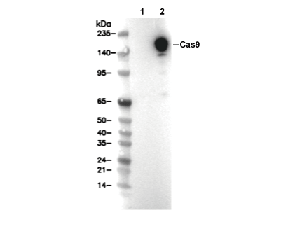 Lane 1: 293T, Lane 2: 293T (transfected with Cas9)