|
Comment citer 1. Pour la citation dans le texte (Matériel & Méthodes) : 2. Pour le tableau des ressources clés : |
||
|
Numéro vert : (877) 796-6397 -- États-Unis et Canada uniquement -- |
Fax : +1-832-582-8590 Commandes : +1-832-582-8158 |
Support technique : +1-832-582-8158 Ext:3 Veuillez indiquer votre numéro de commande dans le-mail. Nous nous efforçons de répondre à toutes les demandes par e-mail dans un délai dun jour ouvrable. |
Description biologique
| Spécificité | CaV1.3 Antibody [G19D14] détecte les niveaux endogènes de la protéine CaV1.3 totale. |
|---|---|
| Contexte | Le couplage excitation-contraction cardiaque fait référence à la séquence d'événements au cours desquels la stimulation électrique d'un cardiomyocyte déclenche la contraction musculaire dans le cœur. Les canaux Ca²⁺ de type L sont essentiels à ce processus, car ils facilitent l'influx de calcium et contribuent à l'excitabilité membranaire. Quatre sous-types de canaux Ca²⁺ de type L ont été identifiés : Cav1.1, Cav1.2, Cav1.3 et Cav1.4. Cav1.1 se trouve principalement dans les muscles squelettiques, tandis que Cav1.4 est principalement exprimé dans la rétine et certaines cellules immunitaires. Cav1.3 est présent dans le cœur, les régions somatodendritiques des neurones, les cellules endocrines et les cellules sensorielles. Dans le cœur, l'activité de Cav1.3 est modulée par de multiples neurotransmetteurs. La phosphorylation par la protéine kinase A (PKA) dépendante de l'AMPc se produit aux résidus sérine 1743 et 1816 dans la région C-terminale. La protéine kinase C (PKC) régule également Cav1.3 de manière isozyme-spécifique via la phosphorylation de la sérine 81 dans le domaine N-terminal. De plus, l'épissage alternatif au sein de l'extrémité C-terminale influence le comportement du canal, notamment en diminuant l'inactivation dépendante du Ca²⁺. Fonctionnellement, Cav1.3 contribue à la régulation du rythme cardiaque et à la conduction atrioventriculaire (AV). Un dysfonctionnement de Cav1.3 a été associé à des anomalies du nœud sinusal et du nœud AV, ainsi qu'au développement de la fibrillation auriculaire. |
Informations dutilisation
| Application | IHC, FCM | Dilution |
|
||
|---|---|---|---|---|---|
| Réactivité | Human, Mouse | ||||
| Source | Mouse Monoclonal Antibody | MW | |||
| Tampon de stockage | PBS, pH 7.2+50% Glycerol+0.05% BSA+0.01% NaN3 | Stockage (À partir de la date de réception) |
-20°C (avoid freeze-thaw cycles), 2 years | ||
| IHC |
Experimental Protocol:
Deparaffinization/Rehydration
1. Deparaffinize/hydrate sections:
2. Incubate sections in three washes of xylene for 5 min each.
3. Incubate sections in two washes of 100% ethanol for 10 min each.
4. Incubate sections in two washes of 95% ethanol for 10 min each.
5. Wash sections two times in dH2O for 5 min each.
6.Antigen retrieval: For Citrate: Heat slides in a microwave submersed in 1X citrate unmasking solution until boiling is initiated; continue with 10 min at a sub-boiling temperature (95°-98°C). Cool slides on bench top for 30 min.
Staining
1. Wash sections in dH2O three times for 5 min each.
2. Incubate sections in 3% hydrogen peroxide for 10 min.
3. Wash sections in dH2O two times for 5 min each.
4. Wash sections in wash buffer for 5 min.
5. Block each section with 100–400 µl of blocking solution for 1 hr at room temperature.
6. Remove blocking solution and add 100–400 µl primary antibody diluent in to each section. Incubate overnight at 4°C.
7. Remove antibody solution and wash sections with wash buffer three times for 5 min each.
8. Cover section with 1–3 drops HRPas needed. Incubate in a humidified chamber for 30 min at room temperature.
9. Wash sections three times with wash buffer for 5 min each.
10. Add DAB Chromogen Concentrate to DAB Diluent and mix well before use.
11. Apply 100–400 µl DAB to each section and monitor closely. 1–10 min generally provides an acceptable staining intensity.
12. Immerse slides in dH2O.
13. If desired, counterstain sections with hematoxylin.
14. Wash sections in dH2O two times for 5 min each.
15. Dehydrate sections: Incubate sections in 95% ethanol two times for 10 sec each; Repeat in 100% ethanol, incubating sections two times for 10 sec each; Repeat in xylene, incubating sections two times for 10 sec each.
16. Mount sections with coverslips and mounting medium.
|
Références
|
Données dapplication
IHC
Validé par Selleck
-
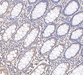 Immunohistochemical analysis of formalin fixed paraffin embedded human colorectal cancer tissue with F3798 at 1:1000 dilution.