|
Comment citer 1. Pour la citation dans le texte (Matériel & Méthodes) : 2. Pour le tableau des ressources clés : |
||
|
Numéro vert : (877) 796-6397 -- États-Unis et Canada uniquement -- |
Fax : +1-832-582-8590 Commandes : +1-832-582-8158 |
Support technique : +1-832-582-8158 Ext:3 Veuillez indiquer votre numéro de commande dans le-mail. Nous nous efforçons de répondre à toutes les demandes par e-mail dans un délai dun jour ouvrable. |
Description biologique
| Spécificité | Collagen IV Antibody [E5G18] détecte les niveaux endogènes de la protéine Collagen IV totale. |
|---|---|
| Contexte | Le Collagen IV est le collagène structural primaire des membranes basales, des matrices extracellulaires spécialisées qui fournissent un support mécanique, régulent l'adhésion cellulaire et organisent d'autres composants de la matrice. Il s'agit d'un hétérotrimère composé de combinaisons de six chaînes α (α1–α6) codées par des gènes distincts, généralement agencées sous forme de deux chaînes α1 et d'une chaîne α2 dans la plupart des tissus, assemblées intracellulairement en protomères triple-hélicoïdaux. Ces protomères sont sécrétés et réticulés via leurs domaines NC1 et 7S pour former un réseau stable, en forme de feuille, qui ancre les laminines, les protéoglycanes et les facteurs de croissance. Le Collagen IV est largement exprimé dans les membranes basales épithéliales et endothéliales, avec une composition d'isoformes spécifique aux tissus, et joue un rôle essentiel dans le développement, l'intégrité tissulaire, la migration cellulaire et la signalisation via des Integrin tels que α1β1 et α2β1. Les défauts ou la réticulation aberrante du Collagen IV contribuent à diverses pathologies, notamment le syndrome d'Alport, la maladie de Goodpasture, la néphropathie diabétique et certaines affections dermatologiques. |
Informations dutilisation
| Application | IHC | Dilution |
|
||
|---|---|---|---|---|---|
| Réactivité | Human | ||||
| Source | Mouse Monoclonal Antibody | MW | |||
| Tampon de stockage | PBS, pH 7.2+50% Glycerol+0.05% BSA+0.01% NaN3 | Stockage (À partir de la date de réception) |
-20°C (avoid freeze-thaw cycles), 2 years | ||
| IHC |
Experimental Protocol:
Deparaffinization/Rehydration
1. Deparaffinize/hydrate sections:
2. Incubate sections in three washes of xylene for 5 min each.
3. Incubate sections in two washes of 100% ethanol for 10 min each.
4. Incubate sections in two washes of 95% ethanol for 10 min each.
5. Wash sections two times in dH2O for 5 min each.
6.Antigen retrieval: For Citrate: Heat slides in a microwave submersed in 1X citrate unmasking solution until boiling is initiated; continue with 10 min at a sub-boiling temperature (95°-98°C). Cool slides on bench top for 30 min.
Staining
1. Wash sections in dH2O three times for 5 min each.
2. Incubate sections in 3% hydrogen peroxide for 10 min.
3. Wash sections in dH2O two times for 5 min each.
4. Wash sections in wash buffer for 5 min.
5. Block each section with 100–400 µl of blocking solution for 1 hr at room temperature.
6. Remove blocking solution and add 100–400 µl primary antibody diluent in to each section. Incubate overnight at 4°C.
7. Remove antibody solution and wash sections with wash buffer three times for 5 min each.
8. Cover section with 1–3 drops HRPas needed. Incubate in a humidified chamber for 30 min at room temperature.
9. Wash sections three times with wash buffer for 5 min each.
10. Add DAB Chromogen Concentrate to DAB Diluent and mix well before use.
11. Apply 100–400 µl DAB to each section and monitor closely. 1–10 min generally provides an acceptable staining intensity.
12. Immerse slides in dH2O.
13. If desired, counterstain sections with hematoxylin.
14. Wash sections in dH2O two times for 5 min each.
15. Dehydrate sections: Incubate sections in 95% ethanol two times for 10 sec each; Repeat in 100% ethanol, incubating sections two times for 10 sec each; Repeat in xylene, incubating sections two times for 10 sec each.
16. Mount sections with coverslips and mounting medium.
|
Références
|
Données dapplication
IHC
Validé par Selleck
-
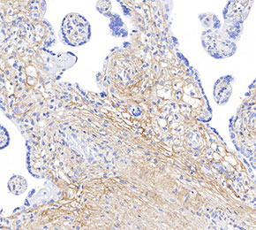 Immunohistochemical analysis of formalin fixed paraffin embedded human placenta tissue with F1667 at 1:125 dilution.