|
Comment citer 1. Pour la citation dans le texte (Matériel & Méthodes) : 2. Pour le tableau des ressources clés : |
||
|
Numéro vert : (877) 796-6397 -- États-Unis et Canada uniquement -- |
Fax : +1-832-582-8590 Commandes : +1-832-582-8158 |
Support technique : +1-832-582-8158 Ext:3 Veuillez indiquer votre numéro de commande dans le-mail. Nous nous efforçons de répondre à toutes les demandes par e-mail dans un délai dun jour ouvrable. |
Description biologique
| Spécificité | CSNK1A1 + CSNK1A1L Antibody [M11N18] détecte les niveaux endogènes de protéines CSNK1A1 et CSNK1A1L totales. |
|---|---|
| Contexte | La caséine kinase 1 alpha 1 (CSNK1A1) et son isoforme CSNK1A1L appartiennent à la famille des caséines kinases 1 des sérine/thréonine kinases, caractérisées par leur préférence pour la phosphorylation de substrats acides tels que les caséines. Elles possèdent un domaine kinase conservé avec des sites de liaison à l'ATP, des motifs de reconnaissance de substrat et des résidus critiques comme le motif DFG et la méthionine « gatekeeper », essentiels pour l'activité enzymatique et la régulation. CSNK1A1 et CSNK1A1L phosphorylent de nombreuses protéines, y compris la β-caténine au niveau de la Ser-45, modulant sa stabilité et jouant ainsi un rôle clé dans les voies de signalisation canoniques Wnt qui régissent la prolifération et la différenciation cellulaires. CSNK1A1 régule les protéines du rythme circadien PER1 et PER2, influence la ségrégation chromosomique pendant la mitose et participe au désassemblage du cytosquelette de kératine, influençant la migration des cellules épithéliales. CSNK1A1 agit comme un régulateur positif des complexes mTOR 1 et 2 (mTORC1/2) en phosphorylant DEPTOR, intégrant les signaux nutritionnels dans les réponses de croissance et métaboliques. Des mutations de novo au sein du domaine kinase de CSNK1A1 affectent les interactions avec le substrat et la régulation en aval de la β-caténine, ce qui est corrélé avec des troubles neurodéveloppementaux. |
Informations dutilisation
| Application | WB | Dilution |
|
||
|---|---|---|---|---|---|
| Réactivité | Mouse, Rat, Human | ||||
| Source | Rabbit Monoclonal Antibody | MW | 39 kDa | ||
| Tampon de stockage | PBS, pH 7.2+50% Glycerol+0.05% BSA+0.01% NaN3 | Stockage (À partir de la date de réception) |
-20°C (avoid freeze-thaw cycles), 2 years | ||
| WB |
Experimental Protocol:
Sample preparation
1. Tissue: Lyse the tissue sample by adding an appropriate volume of ice-cold RIPA/NP-40 Lysis Buffer (containing Protease Inhibitor Cocktail),and homogenize the tissue at a low temperature. 2. Adherent cell: Aspirate the culture medium and wash the cells with ice-cold PBS twice. Lyse the cells by adding an appropriate volume of RIPA/NP-40 Lysis Buffer (containing Protease Inhibitor Cocktail) and put the sample on ice for 5 min. 3. Suspension cell: Transfer the culture medium to a pre-cooled centrifuge tube. Centrifuge and aspirate the supernatant. Wash the cells with ice-cold PBS twice. Lyse the cells by adding an appropriate volume of RIPA/NP-40 Lysis Buffer (containing Protease Inhibitor Cocktail) and put the sample on ice for 5 min. 4. Place the lysate into a pre-cooled microcentrifuge tube. Centrifuge at 4°C for 15 min. Collect the supernatant;
5. Remove a small volume of lysate to determine the protein concentration;
6. Combine the lysate with protein loading buffer. Boil 20 µL sample under 95-100°C for 5 min. Centrifuge for 5 min after cool down on ice.
Electrophoretic separation
1. According to the concentration of extracted protein, load appropriate amount of protein sample and marker onto SDS-PAGE gels for electrophoresis. Recommended separating gel (lower gel) concentration: 10%. Reference Table for Selecting SDS-PAGE Separation Gel Concentrations 2. Power up 80V for 30 minutes. Then the power supply is adjusted (110 V~150 V), the Marker is observed, and the electrophoresis can be stopped when the indicator band of the predyed protein Marker where the protein is located is properly separated. (Note that the current should not be too large when electrophoresis, too large current (more than 150 mA) will cause the temperature to rise, affecting the result of running glue. If high currents cannot be avoided, an ice bath can be used to cool the bath.)
Transfer membrane
1. Take out the converter, soak the clip and consumables in the pre-cooled converter;
2. Activate PVDF membrane with methanol for 1 min and rinse with transfer buffer;
3. Install it in the order of "black edge of clip - sponge - filter paper - filter paper - glue -PVDF membrane - filter paper - filter paper - sponge - white edge of clip"; 4. The protein was electrotransferred to PVDF membrane. ( 0.45 µm PVDF membrane is recommended ) Reference Table for Selecting PVDF Membrane Pore Size Specifications Recommended conditions for wet transfer: 200 mA, 60 min. ( Note that the transfer conditions can be adjusted according to the protein size. For high-molecular-weight proteins, a higher current and longer transfer time are recommended. However, ensure that the transfer tank remains at a low temperature to prevent gel melting.)
Block
1. After electrotransfer, wash the film with TBST at room temperature for 5 minutes;
2. Incubate the film in the blocking solution for 1 hour at room temperature;
3. Wash the film with TBST for 3 times, 5 minutes each time.
Antibody incubation
1. Use 5% skim milk powder to prepare the primary antibody working liquid (recommended dilution ratio for primary antibody 1:1000), gently shake and incubate with the film at 4°C overnight; 2. Wash the film with TBST 3 times, 5 minutes each time;
3. Add the secondary antibody to the blocking solution and incubate with the film gently at room temperature for 1 hour;
4. After incubation, wash the film with TBST 3 times for 5 minutes each time.
Antibody staining
1. Add the prepared ECL luminescent substrate (or select other color developing substrate according to the second antibody) and mix evenly;
2. Incubate with the film for 1 minute, remove excess substrate (keep the film moist), wrap with plastic film, and expose in the imaging system.
|
Références
|
Données dapplication
WB
Validé par Selleck
-
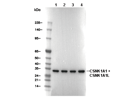 Lane 1: MCF7, Lane 2: HeLa, Lane 3: Jurkat, Lane 4: PC-12