|
Comment citer 1. Pour la citation dans le texte (Matériel & Méthodes) : 2. Pour le tableau des ressources clés : |
||
|
Numéro vert : (877) 796-6397 -- États-Unis et Canada uniquement -- |
Fax : +1-832-582-8590 Commandes : +1-832-582-8158 |
Support technique : +1-832-582-8158 Ext:3 Veuillez indiquer votre numéro de commande dans le-mail. Nous nous efforçons de répondre à toutes les demandes par e-mail dans un délai dun jour ouvrable. |
Description biologique
| Spécificité | GRK 4/5/6 Antibody [P12B19] détecte les niveaux endogènes de la protéine GRK 4/5/6 totale. |
|---|---|
| Contexte | Les récepteurs couplés aux protéines G (GPCRs) constituent une grande famille de protéines membranaires qui médient les réponses cellulaires à un large éventail de ligands extracellulaires par l'activation de divers systèmes de seconds messagers intracellulaires. La terminaison de la signalisation des récepteurs, en particulier la désensibilisation homologue, est principalement contrôlée par les G protein-coupled receptor kinases (GRKs) et leur cofacteur les β-arrestines. Les GRKs phosphorylent les récepteurs occupés par un ligand, facilitant ainsi le recrutement des β-arrestines et favorisant l'internalisation des récepteurs et l'atténuation du signal. La famille des GRK comprend six isoformes (GRK1–6), chacune avec des propriétés régulatrices et une distribution tissulaire distinctes. Parmi elles, GRK4 présente des caractéristiques uniques : elle existe en quatre variants d'épissage alternatif (GRK4α, GRK4β, GRK4γ et GRK4δ) et, contrairement à GRK2, GRK3, GRK5 et GRK6, qui sont largement exprimées dans les tissus, l'expression de GRK4 est fortement restreinte, avec une abondance de transcrits substantielle rapportée principalement dans les testicules. En revanche, GRK6 est plus largement distribuée, en particulier dans le système immunitaire, où elle joue un rôle central dans la modulation des réponses médiées par les GPCR. GRK6 phosphoryle les récepteurs de chimiokines et d'autres GPCR associés au système immunitaire, régulant ainsi la désensibilisation des récepteurs, le trafic et la signalisation en aval. Ses fonctions s'étendent au contrôle des processus inflammatoires, de la migration des leucocytes et de la signalisation nociceptive. Un dérèglement de GRK6 a été impliqué dans les maladies inflammatoires chroniques, y compris la polyarthrite rhumatoïde et les maladies inflammatoires de l'intestin. Sur le plan mécanistique, GRK6 intersecte avec des réseaux transcriptionnels tels que NF-κB, influence la production de cytokines, module la signalisation des espèces réactives de l'oxygène et contribue au maintien des cellules souches hématopoïétiques. |
Informations dutilisation
| Application | WB, IP | Dilution |
|
|---|---|---|---|
| Réactivité | Human, Mouse, Rat | ||
| Source | Mouse Monoclonal Antibody | MW | 65-70kDa |
| Tampon de stockage | PBS, pH 7.2+50% Glycerol+0.05% BSA+0.01% NaN3 | Stockage (À partir de la date de réception) |
-20°C (avoid freeze-thaw cycles), 2 years |
| WB |
Experimental Protocol:
Sample preparation
1. Tissue: Lyse the tissue sample by adding an appropriate volume of ice-cold RIPA/NP-40 Lysis Buffer (containing Protease Inhibitor Cocktail),and homogenize the tissue at a low temperature. 2. Adherent cell: Aspirate the culture medium and wash the cells with ice-cold PBS twice. Lyse the cells by adding an appropriate volume of RIPA/NP-40 Lysis Buffer (containing Protease Inhibitor Cocktail) and put the sample on ice for 5 min. 3. Suspension cell: Transfer the culture medium to a pre-cooled centrifuge tube. Centrifuge and aspirate the supernatant. Wash the cells with ice-cold PBS twice. Lyse the cells by adding an appropriate volume of RIPA/NP-40 Lysis Buffer (containing Protease Inhibitor Cocktail) and put the sample on ice for 5 min. 4. Place the lysate into a pre-cooled microcentrifuge tube. Centrifuge at 4°C for 15 min. Collect the supernatant;
5. Remove a small volume of lysate to determine the protein concentration;
6. Combine the lysate with protein loading buffer. Boil 20 µL sample under 95-100°C for 5 min. Centrifuge for 5 min after cool down on ice.
Electrophoretic separation
1. According to the concentration of extracted protein, load appropriate amount of protein sample and marker onto SDS-PAGE gels for electrophoresis. Recommended separating gel (lower gel) concentration: 10%. Reference Table for Selecting SDS-PAGE Separation Gel Concentrations 2. Power up 80V for 30 minutes. Then the power supply is adjusted (110 V~150 V), the Marker is observed, and the electrophoresis can be stopped when the indicator band of the predyed protein Marker where the protein is located is properly separated. (Note that the current should not be too large when electrophoresis, too large current (more than 150 mA) will cause the temperature to rise, affecting the result of running glue. If high currents cannot be avoided, an ice bath can be used to cool the bath.)
Transfer membrane
1. Take out the converter, soak the clip and consumables in the pre-cooled converter;
2. Activate PVDF membrane with methanol for 1 min and rinse with transfer buffer;
3. Install it in the order of "black edge of clip - sponge - filter paper - filter paper - glue -PVDF membrane - filter paper - filter paper - sponge - white edge of clip"; 4. The protein was electrotransferred to PVDF membrane. ( 0.45 µm PVDF membrane is recommended ) Reference Table for Selecting PVDF Membrane Pore Size Specifications Recommended conditions for wet transfer: 200 mA, 120 min. ( Note that the transfer conditions can be adjusted according to the protein size. For high-molecular-weight proteins, a higher current and longer transfer time are recommended. However, ensure that the transfer tank remains at a low temperature to prevent gel melting.)
Block
1. After electrotransfer, wash the film with TBST at room temperature for 5 minutes;
2. Incubate the film in the blocking solution for 1 hour at room temperature;
3. Wash the film with TBST for 3 times, 5 minutes each time.
Antibody incubation
1. Use 5% skim milk powder to prepare the primary antibody working liquid (recommended dilution ratio for primary antibody 1:500), gently shake and incubate with the film at 4°C overnight; 2. Wash the film with TBST 3 times, 5 minutes each time;
3. Add the secondary antibody to the blocking solution and incubate with the film gently at room temperature for 1 hour;
4. After incubation, wash the film with TBST 3 times for 5 minutes each time.
Antibody staining
1. Add the prepared ECL luminescent substrate (or select other color developing substrate according to the second antibody) and mix evenly;
2. Incubate with the film for 1 minute, remove excess substrate (keep the film moist), wrap with plastic film, and expose in the imaging system.
|
Références
|
Données dapplication
WB
Validé par Selleck
-
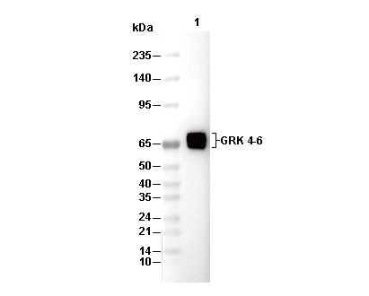 Lane 1: NIH/3T3