|
Comment citer 1. Pour la citation dans le texte (Matériel & Méthodes) : 2. Pour le tableau des ressources clés : |
||
|
Numéro vert : (877) 796-6397 -- États-Unis et Canada uniquement -- |
Fax : +1-832-582-8590 Commandes : +1-832-582-8158 |
Support technique : +1-832-582-8158 Ext:3 Veuillez indiquer votre numéro de commande dans le-mail. Nous nous efforçons de répondre à toutes les demandes par e-mail dans un délai dun jour ouvrable. |
Description biologique
| Spécificité | Histone H4 (tri methyl Lys20) Antibody [J13H2] reconnaît les niveaux endogènes de la protéine Histone H4 uniquement lorsqu'elle est triméthylée en Lys20. Cet anticorps ne réagit pas de manière croisée avec l'Histone H4 Lys20 non méthylée, monométhylée ou diméthylée. Cet anticorps détecte une protéine non spécifique de 95 kDa d'origine inconnue. |
|---|---|
| Contexte | La Tri-Méthyl-Histone H4 (Lys20), également connue sous le nom de H4K20me3, est une modification post-traductionnelle spécifique des histones, cruciale pour le maintien de l'intégrité génomique et la régulation de la structure de la chromatine. La méthylation de la lysine 20 de l'histone H4 existe sous trois états : monométhylation (H4K20me1), diméthylation (H4K20me2) et triméthylation (H4K20me3), H4K20me3 étant le marqueur de triméthylation sur le résidu lysine 20 de l'histone H4. Cette modification est conservée évolutivement de la levure à l'homme et sert de marqueur pour l'hétérochromatine silencieuse transcriptionnellement, indiquant des régions du génome qui ne sont pas activement transcrites. H4K20me3 est fortement enrichie à des emplacements génomiques spécifiques tels que l'hétérochromatine péricentrique, les télomères, les régions soumises à l'empreinte et les éléments répétitifs, jouant un rôle dans le maintien de ces zones dans un état réprimé. Elle est essentielle pour l'intégrité génomique, tant en l'absence qu'en présence de stress génotoxique, faisant partie du réseau plus large de réponse aux dommages de l'ADN (DDR). Les enzymes SUV4-20H1 et SUV4-20H2 médient principalement la triméthylation de H4K20. Les différents états de méthylation de H4K20 changent dynamiquement tout au long du cycle cellulaire. H4K20me3 reste plus stable et montre des changements moins dramatiques par rapport à H4K20me1. H4K20me3 améliore le repliement de la chromatine et joue un rôle structurel dans l'architecture de la chromatine. Une régulation appropriée de la méthylation de H4K20 est vitale pour le maintien de la structure et de la fonction de la chromatine, car des perturbations dans cette régulation entraînent une instabilité génomique et d'autres dysfonctionnements cellulaires. |
Informations dutilisation
| Application | WB, ChIP | Dilution |
|
||||||
|---|---|---|---|---|---|---|---|---|---|
| Réactivité | Human, Mouse, Rat, Monkey, Xenopus, Bovine, Pig | ||||||||
| Source | Rabbit Monoclonal Antibody | MW | 11 Kda | ||||||
| Tampon de stockage | PBS, pH 7.2+50% Glycerol+0.05% BSA+0.01% NaN₃ | Stockage (À partir de la date de réception) |
–20°C (avoid freeze-thaw cycles), 2 years | ||||||
| WB |
Experimental Protocol:
Sample preparation
1. Tissue: Lyse the tissue sample by adding an appropriate volume of ice-cold RIPA/Nuclear Lysis Buffer (containing Protease Inhibitor Cocktail),and homogenize the tissue at a low temperature or lyse it by sonication on ice, then incubate on ice for 30 minutes. 2. Adherent cell: Aspirate the culture medium and transfer the cells into an EP tube. Wash the cells with ice-cold PBS twice. Add an appropriate volume of RIPA/Nuclear Lysis Buffer (containing Protease Inhibitor Cocktail), sonicate to lyse the cells, and incubate on ice for 30 minutes. 3. Suspension cell: Transfer the culture medium to a pre-cooled centrifuge tube. Centrifuge and aspirate the supernatant. Wash the cells with ice-cold PBS twice.Add an appropriate volume of RIPA/Nuclear Lysis Buffer (containing Protease Inhibitor Cocktail), sonicate to lyse the cells, and incubate on ice for 30 minutes. 4. Place the lysate into a pre-cooled microcentrifuge tube. Centrifuge at 4°C for 15 min. Collect the supernatant;
5. Remove a small volume of lysate to determine the protein concentration;
6. Combine the lysate with protein loading buffer. Boil 20 µL sample under 95-100°C for 5 min. Centrifuge for 5 min after cool down on ice.
Electrophoretic separation
1. According to the concentration of extracted protein, load appropriate amount of protein sample and marker onto SDS-PAGE gels for electrophoresis. Recommended separating gel (lower gel) concentration: 20%. Reference Table for Selecting SDS-PAGE Separation Gel Concentrations 2. Power up 80V for 30 minutes. Then the power supply is adjusted (110 V~150 V), the Marker is observed, and the electrophoresis can be stopped when the indicator band of the predyed protein Marker where the protein is located is properly separated. (Note that the current should not be too large when electrophoresis, too large current (more than 150 mA) will cause the temperature to rise, affecting the result of running glue. If high currents cannot be avoided, an ice bath can be used to cool the bath.)
Transfer membrane
1. Take out the converter, soak the clip and consumables in the pre-cooled converter;
2. Activate PVDF membrane with methanol for 1 min and rinse with transfer buffer;
3. Install it in the order of "black edge of clip - sponge - filter paper - filter paper - glue -PVDF membrane - filter paper - filter paper - sponge - white edge of clip"; 4. The protein was electrotransferred to PVDF membrane. ( 0.22 µm PVDF membrane is recommended )) Reference Table for Selecting PVDF Membrane Pore Size Specifications Recommended conditions for wet transfer: 200 mA, 60 min. ( Note that the transfer conditions can be adjusted according to the protein size. For high-molecular-weight proteins, a higher current and longer transfer time are recommended. However, ensure that the transfer tank remains at a low temperature to prevent gel melting.)
Block
1. After electrotransfer, wash the film with TBST at room temperature for 5 minutes;
2. Incubate the film in the blocking solution for 1 hour at room temperature;
3. Wash the film with TBST for 3 times, 5 minutes each time.
Antibody incubation
1. Use 5% skim milk powder to prepare the primary antibody working liquid (recommended dilution ratio for primary antibody 1:1000), gently shake and incubate with the film at 4°C overnight; 2. Wash the film with TBST 3 times, 5 minutes each time;
3. Add the secondary antibody to the blocking solution and incubate with the film gently at room temperature for 1 hour;
4. After incubation, wash the film with TBST 3 times for 5 minutes each time.
Antibody staining
575. Add the prepared ECL luminescent substrate (or select other color developing substrate according to the second antibody) and mix evenly;
2. Incubate with the film for 1 minute, remove excess substrate (keep the film moist), wrap with plastic film, and expose in the imaging system.
|
Références
|
Données dapplication
WB
Validé par Selleck
-
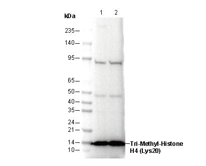 Lane 1: Hela
Lane 2: NIH3T3