|
Comment citer 1. Pour la citation dans le texte (Matériel & Méthodes) : 2. Pour le tableau des ressources clés : |
||
|
Numéro vert : (877) 796-6397 -- États-Unis et Canada uniquement -- |
Fax : +1-832-582-8590 Commandes : +1-832-582-8158 |
Support technique : +1-832-582-8158 Ext:3 Veuillez indiquer votre numéro de commande dans le-mail. Nous nous efforçons de répondre à toutes les demandes par e-mail dans un délai dun jour ouvrable. |
Description biologique
| Spécificité | Lysophospholipase 1 Antibody [F5K18] reconnaît les niveaux endogènes de la protéine totale Lysophospholipase 1/LPL-I. |
|---|---|
| Contexte | La Lysophospholipase 1 (LPL-I), également connue sous le nom de LYPLA1, est une enzyme cytoplasmique essentielle au métabolisme lipidique, hydrolysant principalement les lysophospholipides (LysoPLs), qui sont des intermédiaires clés dans le cycle de Lands responsables du remodelage des phospholipides et de la régulation de la signalisation lipidique. Elle adopte un repliement d'α/β hydrolase avec une triade catalytique Ser-His-Asp conservée qui permet un clivage efficace des chaînes d'acides gras des LysoPLs. LYPLA1 fonctionne également comme une thioestérase qui dépalmitoyle les protéines palmitoylées comme HRAS, régulant ainsi leur localisation subcellulaire, leur association membranaire et leur signalisation. Exprimée de manière ubiquitaire, LYPLA1 aide à maintenir l'homéostasie lipidique cellulaire en prévenant l'accumulation toxique de LysoPLs et en préservant l'intégrité de la membrane. Elle agit de concert avec son isozyme LYPLA2 pour réguler les processus neurodéveloppementaux tels que la différenciation neuronale, en particulier par la modulation de MAPK et d'autres voies de signalisation médiées par les lipides. La dérégulation de LYPLA1 est impliquée dans l'inflammation, la neurodégénérescence et la progression du cancer. Son activité enzymatique est modulée par la disponibilité du substrat et les modifications post-traductionnelles, y compris la palmitoylation, l'acétylation et éventuellement la phosphorylation. |
Informations dutilisation
| Application | WB | Dilution |
|
||
|---|---|---|---|---|---|
| Réactivité | Human, Mouse, Rat | ||||
| Source | Rabbit Monoclonal Antibody | MW | 25 kDa | ||
| Tampon de stockage | PBS, pH 7.2+50% Glycerol+0.05% BSA+0.01% NaN₃ | Stockage (À partir de la date de réception) |
-20°C (avoid freeze-thaw cycles), 2 years | ||
| WB |
Experimental Protocol:
Sample preparation
1. Tissue: Lyse the tissue sample by adding an appropriate volume of ice-cold RIPA/NP-40 Lysis Buffer (containing Protease Inhibitor Cocktail),and homogenize the tissue at a low temperature. 2. Adherent cell: Aspirate the culture medium and wash the cells with ice-cold PBS twice. Lyse the cells by adding an appropriate volume of RIPA/NP-40 Lysis Buffer (containing Protease Inhibitor Cocktail) and put the sample on ice for 5 min. 3. Suspension cell: Transfer the culture medium to a pre-cooled centrifuge tube. Centrifuge and aspirate the supernatant. Wash the cells with ice-cold PBS twice. Lyse the cells by adding an appropriate volume of RIPA/NP-40 Lysis Buffer (containing Protease Inhibitor Cocktail) and put the sample on ice for 5 min. 4. Place the lysate into a pre-cooled microcentrifuge tube. Centrifuge at 4°C for 15 min. Collect the supernatant;
5. Remove a small volume of lysate to determine the protein concentration;
6. Combine the lysate with protein loading buffer. Boil 20 µL sample under 95-100°C for 5 min. Centrifuge for 5 min after cool down on ice.
Electrophoretic separation
1. According to the concentration of extracted protein, load appropriate amount of protein sample and marker onto SDS-PAGE gels for electrophoresis. Recommended separating gel (lower gel) concentration: 10%. Reference Table for Selecting SDS-PAGE Separation Gel Concentrations 2. Power up 80V for 30 minutes. Then the power supply is adjusted (110 V~150 V), the Marker is observed, and the electrophoresis can be stopped when the indicator band of the predyed protein Marker where the protein is located is properly separated. (Note that the current should not be too large when electrophoresis, too large current (more than 150 mA) will cause the temperature to rise, affecting the result of running glue. If high currents cannot be avoided, an ice bath can be used to cool the bath.)
Transfer membrane
1. Take out the converter, soak the clip and consumables in the pre-cooled converter;
2. Activate PVDF membrane with methanol for 1 min and rinse with transfer buffer;
3. Install it in the order of "black edge of clip - sponge - filter paper - filter paper - glue -PVDF membrane - filter paper - filter paper - sponge - white edge of clip"; 4. The protein was electrotransferred to PVDF membrane. ( 0.45 µm PVDF membrane is recommended ) Reference Table for Selecting PVDF Membrane Pore Size Specifications Recommended conditions for wet transfer: 200 mA, 60 min. ( Note that the transfer conditions can be adjusted according to the protein size. For high-molecular-weight proteins, a higher current and longer transfer time are recommended. However, ensure that the transfer tank remains at a low temperature to prevent gel melting.)
Block
1. After electrotransfer, wash the film with TBST at room temperature for 5 minutes;
2. Incubate the film in the blocking solution for 1 hour at room temperature;
3. Wash the film with TBST for 3 times, 5 minutes each time.
Antibody incubation
1. Use 5% skim milk powder to prepare the primary antibody working liquid (recommended dilution ratio for primary antibody 1:5000), gently shake and incubate with the film at 4°C overnight; 2. Wash the film with TBST 3 times, 5 minutes each time;
3. Add the secondary antibody to the blocking solution and incubate with the film gently at room temperature for 1 hour;
4. After incubation, wash the film with TBST 3 times for 5 minutes each time.
Antibody staining
1. Add the prepared ECL luminescent substrate (or select other color developing substrate according to the second antibody) and mix evenly;
2. Incubate with the film for 1 minute, remove excess substrate (keep the film moist), wrap with plastic film, and expose in the imaging system.
|
Références
|
Données dapplication
WB
Validé par Selleck
-
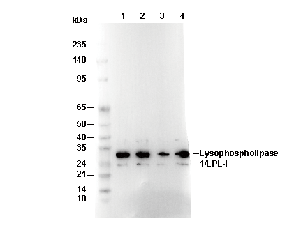 Lane 1: Mouse heart, Lane 2: Mouse kidney, Lane 3: Mouse spleen, Lane 4: Rat kidney