|
Comment citer 1. Pour la citation dans le texte (Matériel & Méthodes) : 2. Pour le tableau des ressources clés : |
||
|
Numéro vert : (877) 796-6397 -- États-Unis et Canada uniquement -- |
Fax : +1-832-582-8590 Commandes : +1-832-582-8158 |
Support technique : +1-832-582-8158 Ext:3 Veuillez indiquer votre numéro de commande dans le-mail. Nous nous efforçons de répondre à toutes les demandes par e-mail dans un délai dun jour ouvrable. |
Description biologique
| Spécificité | MCM3 Antibody [B16A9] reconnaît les niveaux endogènes de la protéine MCM3 totale. |
|---|---|
| Contexte | MCM3 est une sous-unité essentielle du complexe de maintenance des minichromosomes (MCM) 2-7, qui agit comme l'hélicase réplicative essentielle pour l'initiation et l'élongation de la réplication de l'ADN dans les cellules eucaryotes. Cette grande protéine, d'environ 971 acides aminés, partage une homologie significative avec MCM2, en particulier dans trois régions conservées critiques pour l'activité hélicase et la liaison à l'ADN. Structurellement, MCM3 contient de multiples sites de phosphorylation et des interfaces qui médient les interactions avec d'autres sous-unités MCM, notamment MCM5, et est incorporée dans l'anneau hexamérique MCM2-7 qui se charge sur les origines de réplication dans le cadre du complexe pré-réplicatif (pré-RC) pendant les phases M tardive et G1. Le recrutement de MCM3 aux origines dépend du complexe de reconnaissance des origines (ORC), de CDC6 et de CDT1, garantissant que la licence de réplication n'a lieu qu'une seule fois par Cell Cycle. L'activité de MCM3 est étroitement régulée par phosphorylation, notamment par des kinases dépendantes des cyclines telles que la cycline E/Cdk2, qui phosphoryle la thréonine 722 pour promouvoir le chargement de la chromatine mais peut inhiber la réplication en cas de dérégulation. C'est également un substrat pour les kinases de point de contrôle ATM et ATR, liant MCM3 aux réponses aux dommages de l'ADN et aux voies de point de contrôle de la réplication. L'acétylation par MCM3AP inhibe l'initiation de la réplication de l'ADN et la progression du Cell Cycle, ajoutant une autre couche de contrôle. L'isomérase prolyle Pin1 module la liaison de MCM3 à la chromatine de manière dépendante du Cell Cycle, intégrant davantage la régulation post-traductionnelle. La surexpression de MCM3 de type sauvage peut entraîner un arrêt en G1 et un retard de la phase S, tandis que des mutants spécifiques tels que T722A peuvent annuler ces effets. Bien que MCM3 soit essentiel pour la progression de la fourche de réplication et la stabilité du génome, seule une fraction des complexes chargés est activement engagée dans la réplication, les complexes excédentaires servant d'origines de secours et participant à la signalisation des points de contrôle. MCM3 interagit également avec d'autres protéines associées à la réplication et à la chromatine, régulant la spécificité des origines et coordonnant la réplication avec la progression du Cell Cycle. Les mutations ou les déséquilibres dans l'expression de MCM3 peuvent entraîner des défauts dans la licence de réplication, une instabilité génomique et un arrêt du Cell Cycle. |
Informations dutilisation
| Application | WB, IHC, FCM | Dilution |
|
||||||
|---|---|---|---|---|---|---|---|---|---|
| Réactivité | Human, Mouse, Rat | ||||||||
| Source | Rabbit Monoclonal Antibody | MW | 91 kDa | ||||||
| Tampon de stockage | PBS, pH 7.2+50% Glycerol+0.05% BSA+0.01% NaN3 | Stockage (À partir de la date de réception) |
-20°C (avoid freeze-thaw cycles), 2 years | ||||||
| WB |
Experimental Protocol:
Sample preparation
1. Tissue: Lyse the tissue sample by adding an appropriate volume of ice-cold RIPA/Nuclear Lysis Buffer (containing Protease Inhibitor Cocktail),and homogenize the tissue at a low temperature. 2. Adherent cell: Aspirate the culture medium and wash the cells with ice-cold PBS twice. Lyse the cells by adding an appropriate volume of RIPA/Nuclear Lysis Buffer (containing Protease Inhibitor Cocktail) and put the sample on ice for 5 min. 3. Suspension cell: Transfer the culture medium to a pre-cooled centrifuge tube. Centrifuge and aspirate the supernatant. Wash the cells with ice-cold PBS twice. Lyse the cells by adding an appropriate volume of RIPA/Nuclear Lysis Buffer (containing Protease Inhibitor Cocktail) and put the sample on ice for 5 min. 4. Place the lysate into a pre-cooled microcentrifuge tube. Centrifuge at 4°C for 15 min. Collect the supernatant;
5. Remove a small volume of lysate to determine the protein concentration;
6. Combine the lysate with protein loading buffer. Boil 20 µL sample under 95-100°C for 5 min. Centrifuge for 5 min after cool down on ice.
Electrophoretic separation
1. According to the concentration of extracted protein, load appropriate amount of protein sample and marker onto SDS-PAGE gels for electrophoresis. Recommended separating gel (lower gel) concentration: 10%. Reference Table for Selecting SDS-PAGE Separation Gel Concentrations 2. Power up 80V for 30 minutes. Then the power supply is adjusted (110 V~150 V), the Marker is observed, and the electrophoresis can be stopped when the indicator band of the predyed protein Marker where the protein is located is properly separated. (Note that the current should not be too large when electrophoresis, too large current (more than 150 mA) will cause the temperature to rise, affecting the result of running glue. If high currents cannot be avoided, an ice bath can be used to cool the bath.)
Transfer membrane
1. Take out the converter, soak the clip and consumables in the pre-cooled converter;
2. Activate PVDF membrane with methanol for 1 min and rinse with transfer buffer;
3. Install it in the order of "black edge of clip - sponge - filter paper - filter paper - glue -PVDF membrane - filter paper - filter paper - sponge - white edge of clip"; 4. The protein was electrotransferred to PVDF membrane. ( 0.45 µm PVDF membrane is recommended ) Reference Table for Selecting PVDF Membrane Pore Size Specifications Recommended conditions for wet transfer: 200 mA, 120 min. ( Note that the transfer conditions can be adjusted according to the protein size. For high-molecular-weight proteins, a higher current and longer transfer time are recommended. However, ensure that the transfer tank remains at a low temperature to prevent gel melting.)
Block
1. After electrotransfer, wash the film with TBST at room temperature for 5 minutes;
2. Incubate the film in the blocking solution for 1 hour at room temperature;
3. Wash the film with TBST for 3 times, 5 minutes each time.
Antibody incubation
1. Use 5% skim milk powder to prepare the primary antibody working liquid (recommended dilution ratio for primary antibody 1:1000), gently shake and incubate with the film at 4°C overnight; 2. Wash the film with TBST 3 times, 5 minutes each time;
3. Add the secondary antibody to the blocking solution and incubate with the film gently at room temperature for 1 hour;
4. After incubation, wash the film with TBST 3 times for 5 minutes each time.
Antibody staining
1. Add the prepared ECL luminescent substrate (or select other color developing substrate according to the second antibody) and mix evenly;
2. Incubate with the film for 1 minute, remove excess substrate (keep the film moist), wrap with plastic film, and expose in the imaging system.
|
| IHC |
Experimental Protocol:
Deparaffinization/Rehydration
1. Deparaffinize/hydrate sections:
2. Incubate sections in three washes of xylene for 5 min each.
3. Incubate sections in two washes of 100% ethanol for 10 min each.
4. Incubate sections in two washes of 95% ethanol for 10 min each.
5. Wash sections two times in dH2O for 5 min each.
6.Antigen retrieval: For Citrate: Heat slides in a microwave submersed in 1X citrate unmasking solution until boiling is initiated; continue with 10 min at a sub-boiling temperature (95°-98°C). Cool slides on bench top for 30 min.
Staining
1. Wash sections in dH2O three times for 5 min each.
2. Incubate sections in 3% hydrogen peroxide for 10 min.
3. Wash sections in dH2O two times for 5 min each.
4. Wash sections in wash buffer for 5 min.
5. Block each section with 100–400 µl of blocking solution for 1 hr at room temperature.
6. Remove blocking solution and add 100–400 µl primary antibody diluent in to each section. Incubate overnight at 4°C.
7. Remove antibody solution and wash sections with wash buffer three times for 5 min each.
8. Cover section with 1–3 drops HRPas needed. Incubate in a humidified chamber for 30 min at room temperature.
9. Wash sections three times with wash buffer for 5 min each.
10. Add DAB Chromogen Concentrate to DAB Diluent and mix well before use.
11. Apply 100–400 µl DAB to each section and monitor closely. 1–10 min generally provides an acceptable staining intensity.
12. Immerse slides in dH2O.
13. If desired, counterstain sections with hematoxylin.
14. Wash sections in dH2O two times for 5 min each.
15. Dehydrate sections: Incubate sections in 95% ethanol two times for 10 sec each; Repeat in 100% ethanol, incubating sections two times for 10 sec each; Repeat in xylene, incubating sections two times for 10 sec each.
16. Mount sections with coverslips and mounting medium.
|
Références
|
Données dapplication
WB
Validé par Selleck
-
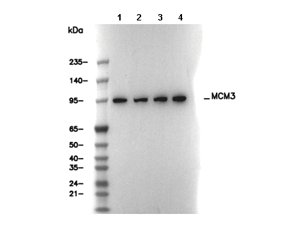 Lane 1: C6, Lane 2: RAW 264.7, Lane 3: PC-12, Lane 4: NIH/3T3