|
Comment citer 1. Pour la citation dans le texte (Matériel & Méthodes) : 2. Pour le tableau des ressources clés : |
||
|
Numéro vert : (877) 796-6397 -- États-Unis et Canada uniquement -- |
Fax : +1-832-582-8590 Commandes : +1-832-582-8158 |
Support technique : +1-832-582-8158 Ext:3 Veuillez indiquer votre numéro de commande dans le-mail. Nous nous efforçons de répondre à toutes les demandes par e-mail dans un délai dun jour ouvrable. |
Description biologique
| Spécificité | Mucin 5AC Antibody [L15D9] détecte les niveaux endogènes de la protéine Mucin 5AC totale. |
|---|---|
| Contexte | La mucine 5AC (MUC5AC) est une glycoprotéine de mucine sécrétée, formant un gel, appartenant à la famille des mucines, principalement exprimée par les cellules caliciformes dans les épithéliums respiratoires, gastriques et conjonctivaux, où elle constitue le composant principal des gels de mucus protecteurs. La MUC5AC est une protéine multidomaine massive présentant des régions globulaires N- et C-terminales avec des domaines D (D1, D2, D2', D3, D4) de type facteur de von Willebrand (VWF), des sous-domaines riches en cystéine (CysD1-9) et un domaine de mucine central riche en proline/thréonine/sérine (PTS) fortement O-glycosylé (>80% de la masse sous forme de glucides) qui s'étend en structures rigides en forme de bâtonnets ; les domaines D1-D3 N-terminaux médient la multimérisation en réseaux étroitement ramifiés via des liaisons disulfures et des interactions homotypiques. La MUC5AC polymérise avec la MUC5B pour former des couches de mucus viscoélastiques qui piègent les agents pathogènes, facilitent la clairance mucociliaire, lubrifient les épithéliums et lient l'eau pour l'hydratation de la barrière ; ses propriétés biophysiques créent des gels plus rigides et plus denses que ceux de la MUC5B en raison de la ramification étendue. L'expression de la MUC5AC est régulée à la hausse via les voies EGFR et IL-13/STAT6 pendant l'inflammation, contribuant à l'obstruction des voies respiratoires dans l'asthme, la BPCO et la mucoviscidose, où l'hypersécrétion altère la clairance ; elle favorise également la progression tumorale dans l'adénocarcinome pulmonaire via la signalisation ANXA2. |
Informations dutilisation
| Application | IHC, IF | Dilution |
|
||||
|---|---|---|---|---|---|---|---|
| Réactivité | Mouse, Rat, Human | ||||||
| Source | Mouse Monoclonal Antibody | MW | |||||
| Tampon de stockage | PBS, pH 7.2+50% Glycerol+0.05% BSA+0.01% NaN3 | Stockage (À partir de la date de réception) |
-20°C (avoid freeze-thaw cycles), 2 years | ||||
| IHC |
Experimental Protocol:
Deparaffinization/Rehydration
1. Deparaffinize/hydrate sections:
2. Incubate sections in three washes of xylene for 5 min each.
3. Incubate sections in two washes of 100% ethanol for 10 min each.
4. Incubate sections in two washes of 95% ethanol for 10 min each.
5. Wash sections two times in dH2O for 5 min each.
6.Antigen retrieval: For Citrate: Heat slides in a microwave submersed in 1X citrate unmasking solution until boiling is initiated; continue with 10 min at a sub-boiling temperature (95°-98°C). Cool slides on bench top for 30 min.
Staining
1. Wash sections in dH2O three times for 5 min each.
2. Incubate sections in 3% hydrogen peroxide for 10 min.
3. Wash sections in dH2O two times for 5 min each.
4. Wash sections in wash buffer for 5 min.
5. Block each section with 100–400 µl of blocking solution for 1 hr at room temperature.
6. Remove blocking solution and add 100–400 µl primary antibody diluent in to each section. Incubate overnight at 4°C.
7. Remove antibody solution and wash sections with wash buffer three times for 5 min each.
8. Cover section with 1–3 drops HRPas needed. Incubate in a humidified chamber for 30 min at room temperature.
9. Wash sections three times with wash buffer for 5 min each.
10. Add DAB Chromogen Concentrate to DAB Diluent and mix well before use.
11. Apply 100–400 µl DAB to each section and monitor closely. 1–10 min generally provides an acceptable staining intensity.
12. Immerse slides in dH2O.
13. If desired, counterstain sections with hematoxylin.
14. Wash sections in dH2O two times for 5 min each.
15. Dehydrate sections: Incubate sections in 95% ethanol two times for 10 sec each; Repeat in 100% ethanol, incubating sections two times for 10 sec each; Repeat in xylene, incubating sections two times for 10 sec each.
16. Mount sections with coverslips and mounting medium.
|
| IF |
Experimental Protocol:
Sample Preparation
1. Adherent Cells: Place a clean, sterile coverslip in a culture dish. Once the cells grow to near confluence as a monolayer, remove the coverslip for further use.
2. Suspension Cells: Seed the cells onto a clean, sterile slide coated with poly-L-lysine.
3. Frozen Sections: Allow the slide to thaw at room temperature. Wash it with pure water or PBS for 2 times, 3 minutes each time.
4. Paraffin Sections: Deparaffinization and rehydration. Wash the slide with pure water or PBS for 3 times, 3 minutes each time. Then perform antigen retrieval.
Fixation
1. Fix the cell coverslips/spots or tissue sections at room temperature using a fixative such as 4% paraformaldehyde (4% PFA) for 10-15 minutes.
2. Wash the sample with PBS for 3 times, 3 minutes each time.
Permeabilization
1.Add a detergent such as 0.1–0.3% Triton X-100 to the sample and incubate at room temperature for 10–20 minutes.
(Note: This step is only required for intracellular antigens. For antigens expressed on the cell membrane, this step is unnecessary.)
Wash the sample with PBS for 3 times, 3 minutes each time.
Blocking
Add blocking solution and incubate at room temperature for at least 1 hour. (Common blocking solutions include: serum from the same source as the secondary antibody, BSA, or goat serum.)
Note: Ensure the sample remains moist during and after the blocking step to prevent drying, which can lead to high background.
Immunofluorescence Staining (Day 1)
1. Remove the blocking solution and add the diluted primary antibody.
2. Incubate the sample in a humidified chamber at 4°C overnight.
Immunofluorescence Staining (Day 2)
1. Remove the primary antibody and wash with PBST for 3 times, 5 minutes each time.
2. Add the diluted fluorescent secondary antibody and incubate in the dark at 4°C for 1–2 hours.
3. Remove the secondary antibody and wash with PBST for 3 times, 5 minutes each time.
4. Add diluted DAPI and incubate at room temperature in the dark for 5–10 minutes.
5. Wash with PBST for 3 times, 5 minutes each time.
Mounting
1. Mount the sample with an anti-fade mounting medium.
2. Allow the slide to dry at room temperature overnight in the dark.
3. Store the slide in a slide storage box at 4°C, protected from light.
|
Références
|
Données dapplication
IF
Validé par Selleck
-
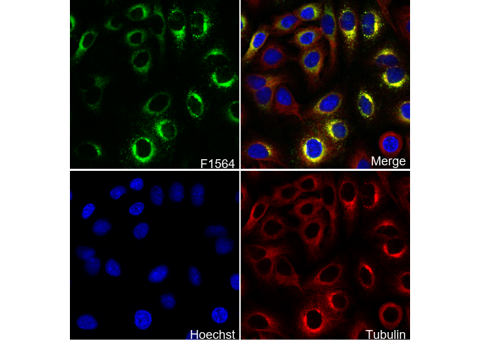 Immunofluorescent analysis of A549 cells using F1564 (green, 1:100), Hoechst (blue) and tubulin (Red).