|
Comment citer 1. Pour la citation dans le texte (Matériel & Méthodes) : 2. Pour le tableau des ressources clés : |
||
|
Numéro vert : (877) 796-6397 -- États-Unis et Canada uniquement -- |
Fax : +1-832-582-8590 Commandes : +1-832-582-8158 |
Support technique : +1-832-582-8158 Ext:3 Veuillez indiquer votre numéro de commande dans le-mail. Nous nous efforçons de répondre à toutes les demandes par e-mail dans un délai dun jour ouvrable. |
Description biologique
| Spécificité | Phospho-IRS-1 (Ser302) Antibody [K1C11] détecte les niveaux endogènes d'IRS-1 uniquement lorsqu'il est phosphorylé sur la Sérine 302. |
|---|---|
| Contexte | La phosphorylation du substrat 1 du récepteur de l'insuline (IRS-1) sur les résidus sérine/thréonine joue un rôle crucial dans la régulation de la signalisation de l'insuline. L'IRS-1 est un substrat primaire de la Protein Tyrosine Kinase du récepteur de l'insuline et contient de multiples sites de phosphorylation de la tyrosine qui agissent comme des plateformes d'ancrage pour les protéines contenant un domaine SH2, facilitant les actions métaboliques et de promotion de la croissance de l'insuline. Les protéines IRS sont caractérisées par de multiples domaines d'interaction et motifs de phosphorylation, mais elles manquent d'activité catalytique intrinsèque. Chaque protéine IRS présente un domaine d'homologie de pleckstrine NH2-terminal, suivi d'un domaine de liaison à la phosphotyrosine, ce qui aide à lier les protéines IRS aux récepteurs activés, tels que ceux de l'insuline, de l'IGF-I et de l'IL-4. L'insuline est essentielle à la régulation métabolique, car elle favorise l'absorption et le stockage des nutriments dans les cellules musculaires et adipeuses tout en supprimant la néoglucogenèse dans le foie. Plusieurs mécanismes peuvent moduler la voie de signalisation de l'insuline, y compris la phosphorylation de la sérine induite par les cytokines, la dégradation des protéines IRS ou l'inhibition directe du récepteur de l'insuline. L'IRS-1 possède plus de 20 sites potentiels de phosphorylation de la sérine qui peuvent être ciblés par diverses kinases. Par exemple, la phosphorylation de la Ser302 est nécessaire à la phosphorylation de la tyrosine de l'IRS-1 induite par l'insuline, reliant potentiellement la signalisation de l'insuline à la disponibilité des nutriments. Pendant ce temps, la phosphorylation de l'IRS-1 au niveau de la Ser1101, médiée par la PKCθ, inhibe la signalisation de l'insuline, offrant une explication potentielle à la résistance à l'insuline observée dans certains modèles d'obésité. |
Informations dutilisation
| Application | WB | Dilution |
|
||
|---|---|---|---|---|---|
| Réactivité | Human, Mouse | ||||
| Source | Rabbit Monoclonal Antibody | MW | 180 kDa | ||
| Tampon de stockage | PBS, pH 7.2+50% Glycerol+0.05% BSA+0.01% NaN₃ | Stockage (À partir de la date de réception) |
-20°C (avoid freeze-thaw cycles), 2 years | ||
| WB |
Experimental Protocol:
Sample preparation
1. Tissue: Lyse the tissue sample by adding an appropriate volume of ice-cold RIPA/NP-40 Lysis Buffer (containing Protease Inhibitor Cocktail, Phosphatase Inhibitor Cocktail),and homogenize the tissue at a low temperature or lyse it by sonication on ice, then incubate on ice for 30 minutes. 2. Adherent cell: Aspirate the culture medium and transfer the cells into an EP tube. Wash the cells with ice-cold PBS twice. Add an appropriate volume of RIPA/NP-40 Lysis Buffer (containing Protease Inhibitor Cocktail, Phosphatase Inhibitor Cocktail), sonicate to lyse the cells, and incubate on ice for 30 minutes. 3. Suspension cell: Transfer the culture medium to a pre-cooled centrifuge tube. Centrifuge and aspirate the supernatant. Wash the cells with ice-cold PBS twice.Add an appropriate volume of RIPA/NP-40 Lysis Buffer (containing Protease Inhibitor Cocktail, Phosphatase Inhibitor Cocktail), sonicate to lyse the cells, and incubate on ice for 30 minutes. 4. Place the lysate into a pre-cooled microcentrifuge tube. Centrifuge at 4°C for 15 min. Collect the supernatant;
5. Remove a small volume of lysate to determine the protein concentration;
6. Combine the lysate with protein loading buffer. Boil 20 µL sample under 95-100°C for 5 min. Centrifuge for 5 min after cool down on ice.
Electrophoretic separation
1. According to the concentration of extracted protein, load appropriate amount of protein sample and marker onto SDS-PAGE gels for electrophoresis. Recommended separating gel (lower gel) concentration: 5%. Reference Table for Selecting SDS-PAGE Separation Gel Concentrations 2. Power up 80V for 30 minutes. Then the power supply is adjusted (110 V~150 V), the Marker is observed, and the electrophoresis can be stopped when the indicator band of the predyed protein Marker where the protein is located is properly separated. (Note that the current should not be too large when electrophoresis, too large current (more than 150 mA) will cause the temperature to rise, affecting the result of running glue. If high currents cannot be avoided, an ice bath can be used to cool the bath.)
Transfer membrane
1. Take out the converter, soak the clip and consumables in the pre-cooled converter;
2. Activate PVDF membrane with methanol for 1 min and rinse with transfer buffer;
3. Install it in the order of "black edge of clip - sponge - filter paper - filter paper - glue -PVDF membrane - filter paper - filter paper - sponge - white edge of clip"; 4. The protein was electrotransferred to PVDF membrane. ( 0.45 µm PVDF membrane is recommended ) Reference Table for Selecting PVDF Membrane Pore Size Specifications Recommended conditions for wet transfer: 200 mA, 120 min. ( Note that the transfer conditions can be adjusted according to the protein size. For high-molecular-weight proteins, a higher current and longer transfer time are recommended. However, ensure that the transfer tank remains at a low temperature to prevent gel melting.)
Block
1. After electrotransfer, wash the film with TBST at room temperature for 5 minutes;
2. Incubate the film in the blocking solution ( recommending 5% BSA solution)
for 1 hour at room temperature;
3. Wash the film with TBST for 3 times, 5 minutes each time.
Antibody incubation
1. Use 5% skim milk powder to prepare the primary antibody working liquid (recommended dilution ratio for primary antibody 1:1000), gently shake and incubate with the film at 4°C overnight; 2. Wash the film with TBST 3 times, 5 minutes each time;
3. Add the secondary antibody to the blocking solution and incubate with the film gently at room temperature for 1 hour;
4. After incubation, wash the film with TBST 3 times for 5 minutes each time.
Antibody staining
1117. Add the prepared ECL luminescent substrate (or select other color developing substrate according to the second antibody) and mix evenly;
2. Incubate with the film for 1 minute, remove excess substrate (keep the film moist), wrap with plastic film, and expose in the imaging system. (Exposure time of at least 90s is recommended)
|
Références
|
Données dapplication
WB
Validé par Selleck
-
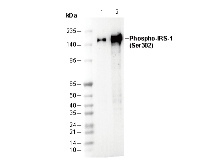 Lane 1: MCF7 (Insulin, 100 nM, 5 min)
Lane 2: C2C12 (Insulin, 100 nM, 5 min)