|
Comment citer 1. Pour la citation dans le texte (Matériel & Méthodes) : 2. Pour le tableau des ressources clés : |
||
|
Numéro vert : (877) 796-6397 -- États-Unis et Canada uniquement -- |
Fax : +1-832-582-8590 Commandes : +1-832-582-8158 |
Support technique : +1-832-582-8158 Ext:3 Veuillez indiquer votre numéro de commande dans le-mail. Nous nous efforçons de répondre à toutes les demandes par e-mail dans un délai dun jour ouvrable. |
Description biologique
| Spécificité | Phospho-STAT1 (Ser727) Antibody [C14E15] reconnaît les niveaux endogènes de STAT1 uniquement lorsqu'il est phosphorylé sur la Sérine 727. |
|---|---|
| Contexte | Phospho-STAT1 (Ser727) fait référence à la phosphorylation de la sérine 727 dans la protéine STAT1, un facteur de transcription clé impliqué dans les réponses cellulaires aux facteurs de stress (par exemple, rayonnement UV, LPS, TNF-α) et la signalisation des cytokines (par exemple, IFN-γ). STAT1, qui fait partie de la famille des Signal Transducer and Activator of Transcription (STAT), contient un domaine N-terminal, un domaine de liaison à l'ADN, un domaine SH2 (pour la dimérisation) et un domaine de transactivation C-terminal (TAD) où réside Ser727. La phosphorylation au niveau de Ser727, médiatisée par des kinases activées par le stress comme p38 MAPK, les ERKs, ou PI-3K/Akt, améliore l'activité transcriptionnelle en recrutant des co-activateurs (par exemple, CBP/p300) et des modificateurs de chromatine, distinctement de la phosphorylation de Tyr701 (voie canonique dépendante de JAK). Cette modification est essentielle pour l'intégration des signaux de stress et immunitaires, amplifiant les réponses antimicrobiennes dans les macrophages et activant les gènes liés à l'apoptose, la survie cellulaire et la mémoire immunitaire. Sous irradiation UVB, la phosphorylation de Ser727 augmente l'affinité de liaison à l'ADN de STAT1 et entraîne des réseaux de gènes réactifs au stress, tandis que dans la signalisation IFN-γ, elle synergise avec la phosphorylation de Tyr701 pour maximiser l'expression des gènes cibles. Une phosphorylation soutenue de Ser727 après le retrait du stimulus (par exemple, IFN-γ) peut maintenir la mémoire transcriptionnelle via des kinases associées à la chromatine comme CDK8. Un dérèglement contribue à l'inflammation, à l'évasion immunitaire et à la carcinogenèse en modulant les gènes suppresseurs de tumeurs et les médiateurs pro-inflammatoires (par exemple, iNOS). |
Informations dutilisation
| Application | WB, IHC | Dilution |
|
||||
|---|---|---|---|---|---|---|---|
| Réactivité | Human, Mouse, Rat | ||||||
| Source | Rabbit Monoclonal Antibody | MW | 87 kDa | ||||
| Tampon de stockage | PBS, pH 7.2+50% Glycerol+0.05% BSA+0.01% NaN3 | Stockage (À partir de la date de réception) |
-20°C (avoid freeze-thaw cycles), 2 years | ||||
| WB |
Experimental Protocol:
Sample preparation
1. Tissue: Lyse the tissue sample by adding an appropriate volume of ice-cold RIPA/NP-40 Lysis Buffer (containing Protease Inhibitor Cocktail),and homogenize the tissue at a low temperature or lyse it by sonication on ice, then incubate on ice for 30 minutes. 2. Adherent cell: Aspirate the culture medium and transfer the cells into an EP tube. Wash the cells with ice-cold PBS twice. Add an appropriate volume of RIPA/NP-40 Lysis Buffer (containing Protease Inhibitor Cocktail), sonicate to lyse the cells, and incubate on ice for 30 minutes. 3. Suspension cell: Transfer the culture medium to a pre-cooled centrifuge tube. Centrifuge and aspirate the supernatant. Wash the cells with ice-cold PBS twice.Add an appropriate volume of RIPA/NP-40 Lysis Buffer (containing Protease Inhibitor Cocktail), sonicate to lyse the cells, and incubate on ice for 30 minutes. 4. Place the lysate into a pre-cooled microcentrifuge tube. Centrifuge at 4°C for 15 min. Collect the supernatant;
5. Remove a small volume of lysate to determine the protein concentration;
6. Combine the lysate with protein loading buffer. Boil 20 µL sample under 95-100°C for 5 min. Centrifuge for 5 min after cool down on ice.
Electrophoretic separation
1. According to the concentration of extracted protein, load appropriate amount of protein sample and marker onto SDS-PAGE gels for electrophoresis. Recommended separating gel (lower gel) concentration: 10%. Reference Table for Selecting SDS-PAGE Separation Gel Concentrations 2. Power up 80V for 30 minutes. Then the power supply is adjusted (110 V~150 V), the Marker is observed, and the electrophoresis can be stopped when the indicator band of the predyed protein Marker where the protein is located is properly separated. (Note that the current should not be too large when electrophoresis, too large current (more than 150 mA) will cause the temperature to rise, affecting the result of running glue. If high currents cannot be avoided, an ice bath can be used to cool the bath.)
Transfer membrane
1. Take out the converter, soak the clip and consumables in the pre-cooled converter;
2. Activate PVDF membrane with methanol for 1 min and rinse with transfer buffer;
3. Install it in the order of "black edge of clip - sponge - filter paper - filter paper - glue -PVDF membrane - filter paper - filter paper - sponge - white edge of clip"; 4. The protein was electrotransferred to PVDF membrane. ( 0.45 µm PVDF membrane is recommended ) Reference Table for Selecting PVDF Membrane Pore Size Specifications Recommended conditions for wet transfer: 200 mA, 120 min. ( Note that the transfer conditions can be adjusted according to the protein size. For high-molecular-weight proteins, a higher current and longer transfer time are recommended. However, ensure that the transfer tank remains at a low temperature to prevent gel melting.)
Block
1. After electrotransfer, wash the film with TBST at room temperature for 5 minutes;
2. Incubate the film in the blocking solution for 1 hour at room temperature;
3. Wash the film with TBST for 3 times, 5 minutes each time.
Antibody incubation
1. Use 5% skim milk powder to prepare the primary antibody working liquid (recommended dilution ratio for primary antibody 1:10000), gently shake and incubate with the film at 4°C overnight; 2. Wash the film with TBST 3 times, 5 minutes each time;
3. Add the secondary antibody to the blocking solution and incubate with the film gently at room temperature for 1 hour;
4. After incubation, wash the film with TBST 3 times for 5 minutes each time.
Antibody staining
1389. Add the prepared ECL luminescent substrate (or select other color developing substrate according to the second antibody) and mix evenly;
2. Incubate with the film for 1 minute, remove excess substrate (keep the film moist), wrap with plastic film, and expose in the imaging system.
|
Références
|
Données dapplication
WB
Validé par Selleck
-
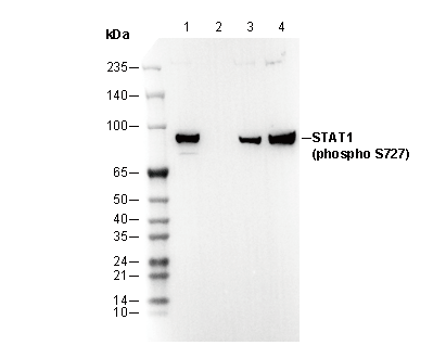 Lane 1: HeLa, Lane 2: HeLa (Alkaline Phosphatase treated), Lane 3: Mouse brain, Lane 4: Rat brain