|
Comment citer 1. Pour la citation dans le texte (Matériel & Méthodes) : 2. Pour le tableau des ressources clés : |
||
|
Numéro vert : (877) 796-6397 -- États-Unis et Canada uniquement -- |
Fax : +1-832-582-8590 Commandes : +1-832-582-8158 |
Support technique : +1-832-582-8158 Ext:3 Veuillez indiquer votre numéro de commande dans le-mail. Nous nous efforçons de répondre à toutes les demandes par e-mail dans un délai dun jour ouvrable. |
Description biologique
| Spécificité | RIG-I / DDX58 Antibody [H1P18] reconnaît les niveaux endogènes de la protéine totale RIG-I / DDX58. |
|---|---|
| Contexte | RIG-I (Retinoic acid-Inducible Gene I), codé par DDX58, est un récepteur de reconnaissance de motifs (PRR) cytosolique essentiel pour la détection de l'ARN viral et le déclenchement de l'immunité innée antivirale. Il est constitué de deux domaines N-terminaux d'activation et de recrutement de la caspase (CARDs), d'un cœur d'hélicase central avec activité ATPase (domaines Hel1 et Hel2), et d'un domaine C-terminal (CTD) qui lie les caractéristiques de l'ARN viral, telles que les extrémités 5'-triphosphate (5'-ppp) ou 5'-diphosphate (5'-pp) et les courts ARN double brin (dsRNA). RIG-I discrimine l'ARN viral de l'ARN de l'hôte par des modifications de l'ARN de l'hôte comme la 2'-O-méthylation, qui empêche l'auto-reconnaissance. Lors de la liaison à l'ARN viral, RIG-I subit des changements conformationnels, exposant ses CARDs pour interagir avec TRIM25, qui ubiquitine RIG-I, et avec MAVS, déclenchant les kinases TBK1/IKKε pour activer IRF3/7 et NF-κB. Cela conduit à la production d'interférons de type I (IFN) et de cytokines pro-inflammatoires. L'hydrolyse de l'ATP du domaine hélicase est cruciale pour le déroulement de l'ARN et l'oligomérisation de RIG-I, favorisant l'agrégation de MAVS pour une signalisation améliorée. De plus, RIG-I détecte les intermédiaires d'ARN dérivés de l'ADN de l'ARN polymérase III, élargissant son rôle dans la détection des virus à ADN. L'activité de RIG-I est régulée par des modifications post-traductionnelles telles que la phosphorylation (par exemple, PKC-α/β) et l'acétylation (par exemple, à K909), qui inhibent sa fonction jusqu'à ce que la déphosphorylation ou la désacétylation médiée par HDAC6 restaure l'activité. Une dérégulation due à des mutations (par exemple, décalage du cadre de lecture DDX58) ou une production aberrante de circRIG-I conduit à des conditions telles que le syndrome de Singleton-Merten et des cancers Immunology & Inflammation related. |
Informations dutilisation
| Application | WB, IP, IF | Dilution |
|
||||||
|---|---|---|---|---|---|---|---|---|---|
| Réactivité | Mouse | ||||||||
| Source | Rabbit Monoclonal Antibody | MW | 102 kDa, 80 kDa | ||||||
| Tampon de stockage | PBS, pH 7.2+50% Glycerol+0.05% BSA+0.01% NaN3 | Stockage (À partir de la date de réception) |
-20°C (avoid freeze-thaw cycles), 2 years | ||||||
| WB |
Experimental Protocol:
Sample preparation
1. Tissue: Lyse the tissue sample by adding an appropriate volume of ice-cold RIPA/NP-40 Lysis Buffer (containing Protease Inhibitor Cocktail),and homogenize the tissue at a low temperature or lyse it by sonication on ice, then incubate on ice for 30 minutes. 2. Adherent cell: Aspirate the culture medium and transfer the cells into an EP tube. Wash the cells with ice-cold PBS twice. Add an appropriate volume of RIPA/NP-40 Lysis Buffer (containing Protease Inhibitor Cocktail), sonicate to lyse the cells, and incubate on ice for 30 minutes. 3. Suspension cell: Transfer the culture medium to a pre-cooled centrifuge tube. Centrifuge and aspirate the supernatant. Wash the cells with ice-cold PBS twice.Add an appropriate volume of RIPA/NP-40 Lysis Buffer (containing Protease Inhibitor Cocktail), sonicate to lyse the cells, and incubate on ice for 30 minutes. 4. Place the lysate into a pre-cooled microcentrifuge tube. Centrifuge at 4°C for 15 min. Collect the supernatant;
5. Remove a small volume of lysate to determine the protein concentration;
6. Combine the lysate with protein loading buffer. Boil 20 µL sample under 95-100°C for 5 min. Centrifuge for 5 min after cool down on ice.
Electrophoretic separation
1. According to the concentration of extracted protein, load appropriate amount of protein sample and marker onto SDS-PAGE gels for electrophoresis. Recommended separating gel (lower gel) concentration: 5%. Reference Table for Selecting SDS-PAGE Separation Gel Concentrations 2. Power up 80V for 30 minutes. Then the power supply is adjusted (110 V~150 V), the Marker is observed, and the electrophoresis can be stopped when the indicator band of the predyed protein Marker where the protein is located is properly separated. (Note that the current should not be too large when electrophoresis, too large current (more than 150 mA) will cause the temperature to rise, affecting the result of running glue. If high currents cannot be avoided, an ice bath can be used to cool the bath.)
Transfer membrane
1. Take out the converter, soak the clip and consumables in the pre-cooled converter;
2. Activate PVDF membrane with methanol for 1 min and rinse with transfer buffer;
3. Install it in the order of "black edge of clip - sponge - filter paper - filter paper - glue -PVDF membrane - filter paper - filter paper - sponge - white edge of clip"; 4. The protein was electrotransferred to PVDF membrane. ( 0.45 µm PVDF membrane is recommended ) Reference Table for Selecting PVDF Membrane Pore Size Specifications Recommended conditions for wet transfer: 200 mA, 120 min. ( Note that the transfer conditions can be adjusted according to the protein size. For high-molecular-weight proteins, a higher current and longer transfer time are recommended. However, ensure that the transfer tank remains at a low temperature to prevent gel melting.)
Block
1. After electrotransfer, wash the film with TBST at room temperature for 5 minutes;
2. Incubate the film in the blocking solution for 1 hour at room temperature;
3. Wash the film with TBST for 3 times, 5 minutes each time.
Antibody incubation
1. Use 5% skim milk powder to prepare the primary antibody working liquid (recommended dilution ratio for primary antibody 1:1000), gently shake and incubate with the film at 4°C overnight; 2. Wash the film with TBST 3 times, 5 minutes each time;
3. Add the secondary antibody to the blocking solution and incubate with the film gently at room temperature for 1 hour;
4. After incubation, wash the film with TBST 3 times for 5 minutes each time.
Antibody staining
1389. Add the prepared ECL luminescent substrate (or select other color developing substrate according to the second antibody) and mix evenly;
2. Incubate with the film for 1 minute, remove excess substrate (keep the film moist), wrap with plastic film, and expose in the imaging system.
|
Références
|
Données dapplication
IF
Validé par Selleck
-
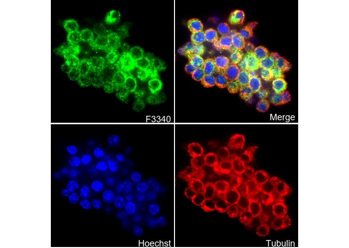 Immunofluorescent analysis of Raw264.7 cells using F3340 (green, 1:500), Hoechst (blue) and tubulin (Red).
WB
Validé par Selleck
-
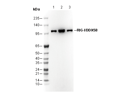 Lane 1: RAW264.7, Lane 2: Mouse spleen, Lane 3: Mouse pancreas