|
Comment citer 1. Pour la citation dans le texte (Matériel & Méthodes) : 2. Pour le tableau des ressources clés : |
||
|
Numéro vert : (877) 796-6397 -- États-Unis et Canada uniquement -- |
Fax : +1-832-582-8590 Commandes : +1-832-582-8158 |
Support technique : +1-832-582-8158 Ext:3 Veuillez indiquer votre numéro de commande dans le-mail. Nous nous efforçons de répondre à toutes les demandes par e-mail dans un délai dun jour ouvrable. |
Description biologique
| Spécificité | S100A9 + Calprotectin (S100A8/A9 complex) Antibody [P14P6] détecte les niveaux endogènes de la protéine totale S100A9 + Calprotectin (S100A8/A9 complex). |
|---|---|
| Contexte | S100A9 + Calprotectin (S100A8/A9 complex), également connu sous le nom de protéines apparentées aux myéloïdes MRP8/MRP14, est un complexe hétérodimère liant le Ca²⁺ composé de S100A8 et S100A9, tous deux membres de la famille des protéines S100. Principalement exprimé dans les neutrophiles, les monocytes et d'autres cellules immunitaires, son expression est induite dans des conditions inflammatoires et de stress. Structurellement, chaque sous-unité contient deux domaines de liaison au Ca²⁺ de type EF-main (un N-terminal à faible affinité et un C-terminal à haute affinité) connectés par une région charnière, formant des hétérodimères stables ou des tétramères fonctionnels. Fonctionnellement, S100A8/A9 joue divers rôles dans l'immunité innée et l'inflammation en régulant la chimiotaxie des leucocytes, la migration des phagocytes, la polymérisation des microtubules, la production d'espèces réactives de l'oxygène et la libération de cytokines. Il se lie également à des récepteurs tels que TLR4 et RAGE, activant des voies de signalisation en aval comme les MAPK, PI3K/Akt, NF-κB et mTOR. Dans le cancer, S100A8/A9 module la croissance tumorale, les métastases, l'apoptose, la résistance aux médicaments et le pronostic, ce qui en fait un biomarqueur et une cible thérapeutique prometteurs. |
Informations dutilisation
| Application | IHC | Dilution |
|
||
|---|---|---|---|---|---|
| Réactivité | Human | ||||
| Source | Mouse Monoclonal Antibody | MW | |||
| Tampon de stockage | PBS, pH 7.2+50% Glycerol+0.05% BSA+0.01% NaN3 | Stockage (À partir de la date de réception) |
-20°C (avoid freeze-thaw cycles), 2 years | ||
| IHC |
Experimental Protocol:
Deparaffinization/Rehydration
1. Deparaffinize/hydrate sections:
2. Incubate sections in three washes of xylene for 5 min each.
3. Incubate sections in two washes of 100% ethanol for 10 min each.
4. Incubate sections in two washes of 95% ethanol for 10 min each.
5. Wash sections two times in dH2O for 5 min each.
6.Antigen retrieval: For Citrate: Heat slides in a microwave submersed in 1X citrate unmasking solution until boiling is initiated; continue with 10 min at a sub-boiling temperature (95°-98°C). Cool slides on bench top for 30 min.
Staining
1. Wash sections in dH2O three times for 5 min each.
2. Incubate sections in 3% hydrogen peroxide for 10 min.
3. Wash sections in dH2O two times for 5 min each.
4. Wash sections in wash buffer for 5 min.
5. Block each section with 100–400 µl of blocking solution for 1 hr at room temperature.
6. Remove blocking solution and add 100–400 µl primary antibody diluent in to each section. Incubate overnight at 4°C.
7. Remove antibody solution and wash sections with wash buffer three times for 5 min each.
8. Cover section with 1–3 drops HRPas needed. Incubate in a humidified chamber for 30 min at room temperature.
9. Wash sections three times with wash buffer for 5 min each.
10. Add DAB Chromogen Concentrate to DAB Diluent and mix well before use.
11. Apply 100–400 µl DAB to each section and monitor closely. 1–10 min generally provides an acceptable staining intensity.
12. Immerse slides in dH2O.
13. If desired, counterstain sections with hematoxylin.
14. Wash sections in dH2O two times for 5 min each.
15. Dehydrate sections: Incubate sections in 95% ethanol two times for 10 sec each; Repeat in 100% ethanol, incubating sections two times for 10 sec each; Repeat in xylene, incubating sections two times for 10 sec each.
16. Mount sections with coverslips and mounting medium.
|
Références
|
Données dapplication
IHC
Validé par Selleck
-
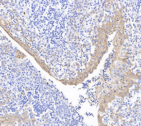 Immunohistochemical analysis of formalin fixed paraffin embedded human tonsils tissue with F1711 at 1:1000 dilution.