|
Comment citer 1. Pour la citation dans le texte (Matériel & Méthodes) : 2. Pour le tableau des ressources clés : |
||
|
Numéro vert : (877) 796-6397 -- États-Unis et Canada uniquement -- |
Fax : +1-832-582-8590 Commandes : +1-832-582-8158 |
Support technique : +1-832-582-8158 Ext:3 Veuillez indiquer votre numéro de commande dans le-mail. Nous nous efforçons de répondre à toutes les demandes par e-mail dans un délai dun jour ouvrable. |
Description biologique
| Spécificité | STAT5 Antibody [B9G7] détecte les niveaux endogènes de la protéine Stat5 totale. Cet anticorps détecte les isoformes alpha et bêta de Stat5 (Stat5a et Stat5b). |
|---|---|
| Contexte | Le transducteur de signal et activateur de transcription 5 (STAT5) comprend deux isoformes hautement homologues, STAT5A et STAT5B, qui fonctionnent comme des médiateurs clés de la signalisation des cytokines et des facteurs de croissance. Ces facteurs de transcription sont activés par les Janus kinases (JAK1, JAK2 et JAK3) après la liaison du ligand aux récepteurs des cytokines. Un large spectre de ligands – y compris l'interleukine-2 (IL-2), le facteur stimulant les colonies de granulocytes et de macrophages (GM-CSF), l'hormone de croissance et la prolactine – peut déclencher l'activation de STAT5. Lors de la stimulation du récepteur, les protéines STAT5 subissent une phosphorylation de la tyrosine, se dimérisent (soit sous forme d'homo- ou d'hétérodimères) et se transloquent vers le noyau, où elles régulent la transcription des target gènes. La signalisation de STAT5 est fondamentale pour le contrôle de divers processus biologiques, y compris la prolifération cellulaire, la différenciation, la survie et les fonctions spécifiques aux tissus telles que l'hématopoïèse et le métabolisme des hépatocytes. L'activité de STAT5 doit être précisément régulée : l'hyperactivation contribue à des états pathologiques tels que le cancer, les troubles myéloprolifératifs, l'inflammation chronique et les maladies auto-immunes, tandis qu'une activation insuffisante peut entraîner une anémie, une thrombocytopénie, un nanisme, une infertilité, une immunodéficience et un dysfonctionnement métabolique. Les deux STAT5A et STAT5B existent en plusieurs isoformes plus courtes générées par épissage alternatif et traitement post-traductionnel. Ces protéines sont en outre modulées par phosphorylation (sur les résidus sérine, thréonine ou tyrosine), ubiquitination et glycosylation, ce qui influence leur stabilité, leur capacité de dimérisation et leur production transcriptionnelle. Par la formation de complexes dimériques distincts, STAT5 orchestre des programmes d'expression génique essentiels au maintien de l'homéostasie cellulaire normale et à la coordination des réponses physiologiques systémiques. |
Informations dutilisation
| Application | WB, IP, ChIP | Dilution |
|
||||||
|---|---|---|---|---|---|---|---|---|---|
| Réactivité | Mouse, Rat, Human | ||||||||
| Source | Rabbit Monoclonal Antibody | MW | 90 kDa | ||||||
| Tampon de stockage | PBS, pH 7.2+50% Glycerol+0.05% BSA+0.01% NaN3 | Stockage (À partir de la date de réception) |
-20°C (avoid freeze-thaw cycles), 2 years | ||||||
| WB |
Experimental Protocol:
Sample preparation
1. Tissue: Lyse the tissue sample by adding an appropriate volume of ice-cold RIPA/NP-40 Lysis Buffer (containing Protease Inhibitor Cocktail),and homogenize the tissue at a low temperature. 2. Adherent cell: Aspirate the culture medium and wash the cells with ice-cold PBS twice. Lyse the cells by adding an appropriate volume of RIPA/NP-40 Lysis Buffer (containing Protease Inhibitor Cocktail) and put the sample on ice for 5 min. 3. Suspension cell: Transfer the culture medium to a pre-cooled centrifuge tube. Centrifuge and aspirate the supernatant. Wash the cells with ice-cold PBS twice. Lyse the cells by adding an appropriate volume of RIPA/NP-40 Lysis Buffer (containing Protease Inhibitor Cocktail) and put the sample on ice for 5 min. 4. Place the lysate into a pre-cooled microcentrifuge tube. Centrifuge at 4°C for 15 min. Collect the supernatant;
5. Remove a small volume of lysate to determine the protein concentration;
6. Combine the lysate with protein loading buffer. Boil 20 µL sample under 95-100°C for 5 min. Centrifuge for 5 min after cool down on ice.
Electrophoretic separation
1. According to the concentration of extracted protein, load appropriate amount of protein sample and marker onto SDS-PAGE gels for electrophoresis. Recommended separating gel (lower gel) concentration: 10%. Reference Table for Selecting SDS-PAGE Separation Gel Concentrations 2. Power up 80V for 30 minutes. Then the power supply is adjusted (110 V~150 V), the Marker is observed, and the electrophoresis can be stopped when the indicator band of the predyed protein Marker where the protein is located is properly separated. (Note that the current should not be too large when electrophoresis, too large current (more than 150 mA) will cause the temperature to rise, affecting the result of running glue. If high currents cannot be avoided, an ice bath can be used to cool the bath.)
Transfer membrane
1. Take out the converter, soak the clip and consumables in the pre-cooled converter;
2. Activate PVDF membrane with methanol for 1 min and rinse with transfer buffer;
3. Install it in the order of "black edge of clip - sponge - filter paper - filter paper - glue -PVDF membrane - filter paper - filter paper - sponge - white edge of clip"; 4. The protein was electrotransferred to PVDF membrane. ( 0.45 µm PVDF membrane is recommended ) Reference Table for Selecting PVDF Membrane Pore Size Specifications Recommended conditions for wet transfer: 200 mA, 120 min. ( Note that the transfer conditions can be adjusted according to the protein size. For high-molecular-weight proteins, a higher current and longer transfer time are recommended. However, ensure that the transfer tank remains at a low temperature to prevent gel melting.)
Block
1. After electrotransfer, wash the film with TBST at room temperature for 5 minutes;
2. Incubate the film in the blocking solution for 1 hour at room temperature;
3. Wash the film with TBST for 3 times, 5 minutes each time.
Antibody incubation
1. Use 5% skim milk powder to prepare the primary antibody working liquid (recommended dilution ratio for primary antibody 1:1000), gently shake and incubate with the film at 4°C overnight; 2. Wash the film with TBST 3 times, 5 minutes each time;
3. Add the secondary antibody to the blocking solution and incubate with the film gently at room temperature for 1 hour;
4. After incubation, wash the film with TBST 3 times for 5 minutes each time.
Antibody staining
1. Add the prepared ECL luminescent substrate (or select other color developing substrate according to the second antibody) and mix evenly;
2. Incubate with the film for 1 minute, remove excess substrate (keep the film moist), wrap with plastic film, and expose in the imaging system.
|
Références
|
Données dapplication
WB
Validé par Selleck
-
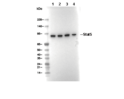 Lane 1: K562, Lane 2: MOLT4, Lane 3: A20, Lane 4: Daudi