|
Comment citer 1. Pour la citation dans le texte (Matériel & Méthodes) : 2. Pour le tableau des ressources clés : |
||
|
Numéro vert : (877) 796-6397 -- États-Unis et Canada uniquement -- |
Fax : +1-832-582-8590 Commandes : +1-832-582-8158 |
Support technique : +1-832-582-8158 Ext:3 Veuillez indiquer votre numéro de commande dans le-mail. Nous nous efforçons de répondre à toutes les demandes par e-mail dans un délai dun jour ouvrable. |
Description biologique
| Spécificité | TR4/NR2C2 Antibody [C17H11] détecte les niveaux endogènes de la protéine TR4/NR2C2 totale. |
|---|---|
| Contexte | Le récepteur testiculaire 4 (TR4), également connu sous le nom de NR2C2 ou TAK1, est un récepteur nucléaire orphelin classé dans la superfamille des récepteurs nucléaires des hormones stéroïdiennes, étroitement lié à TR2. Le TR4 contient les domaines typiques des récepteurs nucléaires, y compris un domaine N-terminal (NTD), un domaine de liaison à l'ADN (DBD) qui reconnaît des éléments de réponse à l'ADN spécifiques souvent sous forme d'homodimères ou d'hétérodimères avec TR2, une région charnière et un domaine de liaison au ligand (LBD) qui peut adopter une conformation auto-réprimée mais est activé par des ligands tels que le rétinol et l'acide rétinoïque. Le TR4 agit comme un facteur de transcription, régulant les gènes impliqués dans les voies métaboliques et reproductives, telles que le métabolisme des lipides et du glucose, la signalisation de l'insuline et les voies du récepteur aux androgènes, souvent en formant des complexes avec d'autres récepteurs nucléaires, tels que l'AR (récepteur aux androgènes) et l'ER (récepteur aux œstrogènes), qui modulent la transcription génique positivement ou négativement. Le TR4 joue un rôle essentiel dans la neurogenèse, la spermatogenèse, le métabolisme de l'ARN et la traduction des protéines et a été lié à des conditions pathologiques, notamment la métastase du cancer de la prostate et le syndrome métabolique. Sa capacité à recruter des coactivateurs ou des corépresseurs, ainsi que des modifications post-traductionnelles telles que la phosphorylation par les MAP kinases, affine son activité transcriptionnelle. L'expression élevée de TR4 est corrélée à un mauvais pronostic dans plusieurs cancers, y compris le cancer de la prostate et le cancer du poumon non à petites cellules. |
Informations dutilisation
| Application | WB, IP | Dilution |
|
||||
|---|---|---|---|---|---|---|---|
| Réactivité | Human, Mouse, Rat, Monkey | ||||||
| Source | Rabbit Monoclonal Antibody | MW | 67 kDa | ||||
| Tampon de stockage | PBS, pH 7.2+50% Glycerol+0.05% BSA+0.01% NaN3 | Stockage (À partir de la date de réception) |
-20°C (avoid freeze-thaw cycles), 2 years | ||||
| WB |
Experimental Protocol:
Sample preparation
1. Tissue: Lyse the tissue sample by adding an appropriate volume of ice-cold RIPA/Nuclear Lysis Buffer (containing Protease Inhibitor Cocktail),and homogenize the tissue at a low temperature. 2. Adherent cell: Aspirate the culture medium and wash the cells with ice-cold PBS twice. Lyse the cells by adding an appropriate volume of RIPA/Nuclear Lysis Buffer (containing Protease Inhibitor Cocktail) and put the sample on ice for 5 min. 3. Suspension cell: Transfer the culture medium to a pre-cooled centrifuge tube. Centrifuge and aspirate the supernatant. Wash the cells with ice-cold PBS twice. Lyse the cells by adding an appropriate volume of RIPA/Nuclear Lysis Buffer (containing Protease Inhibitor Cocktail) and put the sample on ice for 5 min. 4. Place the lysate into a pre-cooled microcentrifuge tube. Centrifuge at 4°C for 15 min. Collect the supernatant;
5. Remove a small volume of lysate to determine the protein concentration;
6. Combine the lysate with protein loading buffer. Boil 20 µL sample under 95-100°C for 5 min. Centrifuge for 5 min after cool down on ice.
Electrophoretic separation
1. According to the concentration of extracted protein, load appropriate amount of protein sample and marker onto SDS-PAGE gels for electrophoresis. Recommended separating gel (lower gel) concentration: 10%. Reference Table for Selecting SDS-PAGE Separation Gel Concentrations 2. Power up 80V for 30 minutes. Then the power supply is adjusted (110 V~150 V), the Marker is observed, and the electrophoresis can be stopped when the indicator band of the predyed protein Marker where the protein is located is properly separated. (Note that the current should not be too large when electrophoresis, too large current (more than 150 mA) will cause the temperature to rise, affecting the result of running glue. If high currents cannot be avoided, an ice bath can be used to cool the bath.)
Transfer membrane
1. Take out the converter, soak the clip and consumables in the pre-cooled converter;
2. Activate PVDF membrane with methanol for 1 min and rinse with transfer buffer;
3. Install it in the order of "black edge of clip - sponge - filter paper - filter paper - glue -PVDF membrane - filter paper - filter paper - sponge - white edge of clip"; 4. The protein was electrotransferred to PVDF membrane. ( 0.45 µm PVDF membrane is recommended ) Reference Table for Selecting PVDF Membrane Pore Size Specifications Recommended conditions for wet transfer: 200 mA, 120 min. ( Note that the transfer conditions can be adjusted according to the protein size. For high-molecular-weight proteins, a higher current and longer transfer time are recommended. However, ensure that the transfer tank remains at a low temperature to prevent gel melting.)
Block
1. After electrotransfer, wash the film with TBST at room temperature for 5 minutes;
2. Incubate the film in the blocking solution for 1 hour at room temperature;
3. Wash the film with TBST for 3 times, 5 minutes each time.
Antibody incubation
1. Use 5% skim milk powder to prepare the primary antibody working liquid (recommended dilution ratio for primary antibody 1:1000), gently shake and incubate with the film at 4°C overnight; 2. Wash the film with TBST 3 times, 5 minutes each time;
3. Add the secondary antibody to the blocking solution and incubate with the film gently at room temperature for 1 hour;
4. After incubation, wash the film with TBST 3 times for 5 minutes each time.
Antibody staining
1. Add the prepared ECL luminescent substrate (or select other color developing substrate according to the second antibody) and mix evenly;
2. Incubate with the film for 1 minute, remove excess substrate (keep the film moist), wrap with plastic film, and expose in the imaging system.
|
Références
|
Données dapplication
WB
Validé par Selleck
-
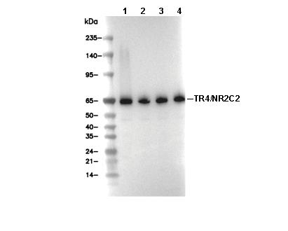 Lane 1: Jurkat, Lane 2: 3T3, Lane 3: C6, Lane 4: COS-7