Usage réservé à la recherche
MCP1 Antibody [M3B19]
N° de cat.: F3225
Application:
Réactivité:
-
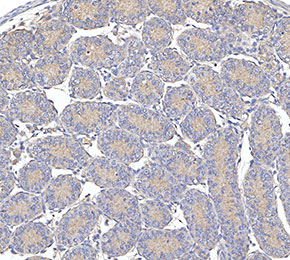 Immunohistochemical analysis of formalin fixed paraffin embedded mouse testicles tissue with F3225 at 1:10 dilution.
Essentiels pour lexpérimentation
This antibody requires an anti-rat secondary antibody.
Informations dutilisation
| Dilution |
|---|
|
|
| Application |
|---|
| IHC |
| Réactivité |
|---|
| Mouse |
| Source |
|---|
| Rat Monoclonal Antibody |
| Tampon de stockage |
|---|
| PBS, pH 7.2+50% Glycerol+0.05% BSA+0.01% NaN3 |
| Stockage (à partir de la date de réception) |
|---|
| -20°C (avoid freeze-thaw cycles), 2 years |
| Poids moléculaire prédit |
|---|
| 11 kDa |
| Contrôle positif | Mouse testis |
|---|---|
| Contrôle négatif |
Méthodes expérimentales
| IHC |
|---|
Experimental Protocol:
Deparaffinization/Rehydration
1. Deparaffinize/hydrate sections:
2. Incubate sections in three washes of xylene for 5 min each.
3. Incubate sections in two washes of 100% ethanol for 10 min each.
4. Incubate sections in two washes of 95% ethanol for 10 min each.
5. Wash sections two times in dH2O for 5 min each.
6.Antigen retrieval: For Citrate: Heat slides in a microwave submersed in 1X citrate unmasking solution until boiling is initiated; continue with 10 min at a sub-boiling temperature (95°-98°C). Cool slides on bench top for 30 min.
Staining
1. Wash sections in dH2O three times for 5 min each.
2. Incubate sections in 3% hydrogen peroxide for 10 min.
3. Wash sections in dH2O two times for 5 min each.
4. Wash sections in wash buffer for 5 min.
5. Block each section with 100–400 µl of blocking solution for 1 hr at room temperature.
6. Remove blocking solution and add 100–400 µl primary antibody diluent in to each section. Incubate overnight at 4°C.
7. Remove antibody solution and wash sections with wash buffer three times for 5 min each.
8. Cover section with 1–3 drops HRPas needed. Incubate in a humidified chamber for 30 min at room temperature.
9. Wash sections three times with wash buffer for 5 min each.
10. Add DAB Chromogen Concentrate to DAB Diluent and mix well before use.
11. Apply 100–400 µl DAB to each section and monitor closely. 1–10 min generally provides an acceptable staining intensity.
12. Immerse slides in dH2O.
13. If desired, counterstain sections with hematoxylin.
14. Wash sections in dH2O two times for 5 min each.
15. Dehydrate sections: Incubate sections in 95% ethanol two times for 10 sec each; Repeat in 100% ethanol, incubating sections two times for 10 sec each; Repeat in xylene, incubating sections two times for 10 sec each.
16. Mount sections with coverslips and mounting medium.
|
Description biologique
| Spécificité |
|---|
MCP1 Antibody [M3B19] detects endogenous levels of total MCP1 protein. |
| Localisation subcellulaire |
|---|
| Secreted |
| Uniprot ID |
|---|
| P10148 |
| Clone |
|---|
| M3B19 |
| Synonyme |
|---|
| Je, Mcp1, Scya2, Ccl2, C-C motif chemokine 2, Monocyte chemoattractant protein 1, Monocyte chemotactic protein 1, Platelet-derived growth factor-inducible protein JE, Small-inducible cytokine A2, MCP-1 |
| Contexte |
|---|
| Monocyte chemoattractant protein-1 (MCP-1/CCL2) is a member of the C-C chemokine family and functions as a potent chemotactic factor for monocytes. It is considered identical to JE, a gene originally identified in mouse fibroblasts as being induced by platelet-derived growth factor. The human MCP-1 gene is located on chromosome 17q11.2 and encodes a protein of 76 amino acids with a molecular weight of approximately 13 kDa. MCP-1 belongs to a subfamily of chemokines that includes at least four members: MCP-1, MCP-2, MCP-3, and MCP-4. CCL2 is produced by a wide variety of cell types, either constitutively or in response to stimuli such as oxidative stress, cytokines, and growth factors. Its sources include endothelial cells, fibroblasts, epithelial cells, smooth muscle cells, mesangial cells, astrocytes, monocytes, and microglia—cell types that play important roles in antiviral immune defense in both circulation and tissues. Functionally, CCL2 directs the migration and infiltration of monocytes, memory T cells, and natural killer (NK) cells. The biological effects of CCL2 are mediated through its receptor, CCR2, whose expression is more restricted compared to that of CCL2. CCR2 exists in two alternatively spliced isoforms, CCR2A and CCR2B, which differ only at their C-terminal regions. |
| Références |
|---|
|
Support technique
Tel: +1-832-582-8158 Ext:3
Si vous avez dautres questions, veuillez laisser un message.
Les produits sont destinés à la recherche uniquement. Ne pas utiliser chez lhomme. Nous ne vendons pas aux patients.
©Copyright 2013 Selleck Chemicals. Tous droits réservés.